【IT1000】EzRNA™ T7 High Yield RNA Synthesis Kit, 50 RXN
Facebook
X
Pinterest
Email
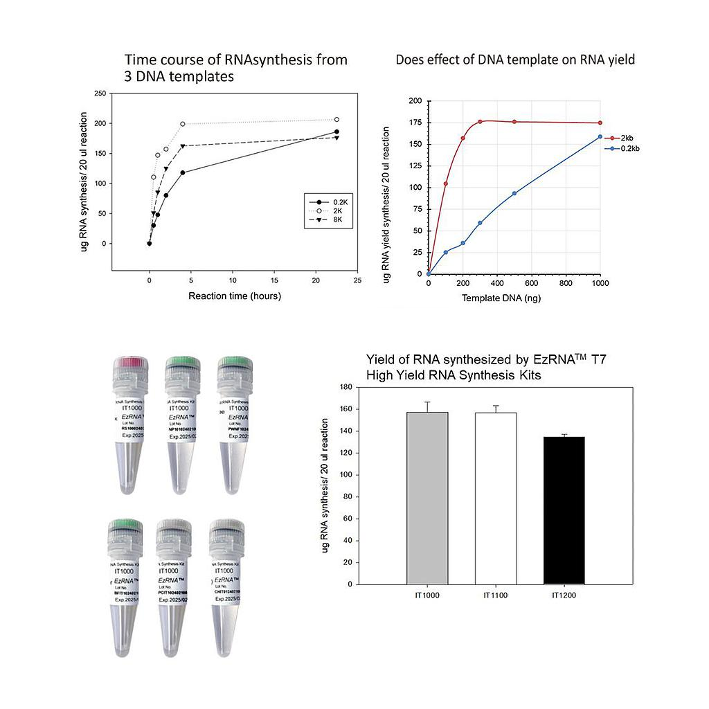
The EzRNA™ T7 High Yield RNA Synthesis Kit is a user-friendly product for enzymatic RNA production. The enzyme mix contains adequate amount of T7 RNA polymerase, pyrophosphatase, and RNase inhibitors for in vitro transcription (IVT). Along with 10X Transcription Buffer and NTP Premix, users can swiftly assemble IVT reactions without compromising RNA yield. The EzRNA™ T7 High Yield RNA Synthesis Kit allows for the attainment of approximately up to 150 µg RNA yield within 2 hours at 37°C.
Detail
Description
The EzRNA™ T7 High Yield RNA Synthesis Kit is a user-friendly product for enzymatic RNA production. The enzyme mix contains adequate amount of T7 RNA polymerase, pyrophosphatase, and RNase inhibitors for in vitro transcription (IVT). Along with 10X Transcription Buffer and NTP Premix, users can swiftly assemble IVT reactions without compromising RNA yield. The EzRNA™ T7 High Yield RNA Synthesis Kit allows for the attainment of approximately up to 150 µg RNA yield within 2 hours at 37°C.
Features
High yield
Versatile- suitable for short and long transcripts
NTP premixed- Minimal pipetting and setup time
Compatible with CleanCap® Reagent AG
Lithium chloride included for RNA purification
Application
Generation of RNA from T7 promoter-driven DNA sequences
Suitable for subsequent cap-0 and cap-1 modification
Organophsphates are a class of pesticides that mechanistically target the acetylcholinesterase enzyme. Regulatory guidelines have been set to ensure our food and water are within the acceptable regulatory authority guidelines. Because most OPs are provided in their precursor form, organothiophosphate (i.e., Malathion, Diazinon, Chlorpyrifos, Azinphos, Dimethoate, Terbufos, Phosmet) Attogene’s organophosphate ELISA kit has been designed to detect organothiophosphates which are the main form of the compounds when applied in the field.
Document
This kit can be used for rapid test of organophosphate in liquid samples such as water, wastewater, and solid samples such as wheat.
96-Well Low Elution Ring Magnetic Plate with Integrated Cushion Base
Product Info
Document
Product Info
Designed for those labs wishing to separate magnetic beads into rings.
The Permagen ring magnet low elution plate with integrated cushion base plate was designed for use in automation applications where volumes as low as 5 µL are required. Most PCR plates are bent, leading to inconsistent lab results. Unlike most products, we have added an angled frame around the top of our plate, this helps with two things, straightening out the PCR plate, and leading it to the proper location on the magnets during automation while the cushion assist compensates for labware inconsistencies.
SBS SLAS Footprint (127.75mm x 85.50mm) to fit into any automated liquid handling robot
Features include solid aluminum alloy construction and hard coat anodized finish for years of trouble-free use, and compatible with any magnetic beads
Designed for those labs wishing to separate magnetic beads into rings.
The Permagen ring magnet low elution plate with integrated cushion base plate was designed for use in automation applications where volumes as low as 5 µL are required. Most PCR plates are bent, leading to inconsistent lab results. Unlike most products, we have added an angled frame around the top of our plate, this helps with two things, straightening out the PCR plate, and leading it to the proper location on the magnets during automation while the cushion assist compensates for labware inconsistencies.
Aspergillus niger Nucleic acid testing (NAT) is the method of choice for detection and quantification of a wide range of micro organisms. Primerdesign manufactures and supplies high quality quantitative real-time PCR kits for the detection and simultaneous quantification of numerous significant pathogens . A copy number standard curve is provided for quantification and an the internal extraction template (DNA or RNA), controls for the quality of the nucleic acid extraction and eliminates false negative results.
The kit is designed with the broadest possible detection profile to ensure that all clinically relevant strains and subtypes are detected. Target sequences are selected by working with data from key opinion leaders in the field. Multiple sequence alignments and unprecedented real-time PCR expertise in design and validation ensure the best possible kit.
Details of the target and priming specificity are included in the individual handbooks above.
Packaged, optimised and ready to use. Expect Better Data.
Document
Exceptional value for money Rapid detection of all clinically relevant subtypes Positive copy number standard curve for quantification Highly specific detection profile High priming efficiency Broad dynamic detection range (>6 logs) Sensitive to < 100 copies of target
Accurate controls to confirm findings