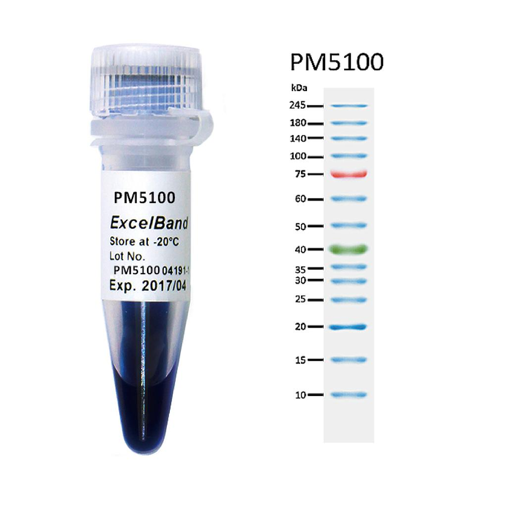
[PM5100] ExcelBand™ 3-color Pre-Stained Protein Ladder, High Range (9-245 kDa), 250 μl x 2
Facebook
X
Pinterest
Email
The PM5100 3-color Pre-Stained Protein Ladder High Range is a ready-to-use three-color protein standard with 14 pre-stained proteins covering a wide range of molecular weights from 10 to 245 kDa in Tris-Glycine Buffer (9 to 235 kDa in Bis-Tris (MOPS) buffer and 10 to 235 kDa in Bis-Tris (MES) buffer). Proteins are covalently coupled with different chromophores for easy identification of bands, with three reference proteins carrying enhanced intensity corresponding to a blue band at 20 kDa, green at 40 kDa, and red at 75 kDa, respectively, as separated on SDS-PAGE (Tris-Glycine buffer). The PM5100 3-color Pre-Stained Protein Ladder High Range is designed for monitoring protein separation during SDS-polyacrylamide gel electrophoresis, verification of Western transfer efficiency on membranes (PVDF, nylon, or nitrocellulose) and for approximating the size of proteins.
Detail
Description
The PM5100 3-color Pre-Stained Protein Ladder High Range is a ready-to-use three-color protein standard with 14 pre-stained proteins covering a wide range of molecular weights from 10 to 245 kDa in Tris-Glycine Buffer (9 to 235 kDa in Bis-Tris (MOPS) buffer and 10 to 235 kDa in Bis-Tris (MES) buffer). Proteins are covalently coupled with different chromophores for easy identification of bands, with three reference proteins carrying enhanced intensity corresponding to a blue band at 20 kDa, green at 40 kDa, and red at 75 kDa, respectively, as separated on SDS-PAGE (Tris-Glycine buffer). The PM5100 3-color Pre-Stained Protein Ladder High Range is designed for monitoring protein separation during SDS-polyacrylamide gel electrophoresis, verification of Western transfer efficiency on membranes (PVDF, nylon, or nitrocellulose) and for approximating the size of proteins.
Features
Ready-to-use — Premixed with a loading buffer for direct loading, no need to boil.
Three reference bands — 75 kDa (red), 40 kDa (green), and 20 kDa (blue)
Contents
Approximately 0.1~0.4 mg/ml of each protein in the buffer (20 mM Tris-phosphate (pH 7.5 at 25°C), 2% SDS, 0.2 mM DTT, 3.6 M urea, and 15% (v/v) glycerol).
Quality Control
Under suggested conditions, PM5100 ExcelBand™ 3-color Pre-Stained Protein Ladder High Range resolves 14 major bands in SDS-PAGE (Tris-Glycine, MOPS, and MES buffer) and after Western blotting to nitrocellulose membrane.
Storage
4°C for 3 months -20°C for long term storage
Other Products
DNA Isothermal Rapid Amplification Kit (Colloidal Gold Test Strip Type)
Product Info
Document
Product Info
Product Details
Application:
DNA Nucleic Acid
Format:
NFO Kit
Kit Components:
Reagents,Buffer,Enzymes
Manufacturer:
Amp-future Bio
Product Name:
DNA Isothermal Rapid Amplification Kit (Colloidal Gold Test Strip Type)
Reaction Time:
5-20mins
Reaction Volume:
50μL
Sensitivity:
500-1000copies/ml
Shelf Life:
14 Months
Specificity:
High
Storage Conditions:
-20℃
Suitability:
Universal
High Light:
Isothermal DNA Amplification Kit
,
DNA Amplification Kit NFO
,
20mins nucleic acid amplification
Payment & Shipping Terms
Minimum Order Quantity
48T
Price
USD$3.8/T
Packaging Details
16T/Bag,48T/Box
Delivery Time
6 working days
Payment Terms
T/T, MoneyGram
Supply Ability
100000T/Month
Product Description
DNA Isothermal Rapid Amplification Kit (Colloidal Gold Test Strip Type)
【Principle Overview】
This kit is based on a rapid nucleic acid amplification technology at room temperature and constant temperature: at room temperature and constant temperature (generally 39℃~42℃), with the help of auxiliary proteins and single-strand binding proteins, the recombinase and primers form a complex; Source search and combine the target homology domain, at this time, a D-loop region is formed at the homology position and strand exchange begins; along with the dissociation of the recombinase from the complex, the polymerase also binds to the 3′ end of the primer and begins chain extension. Relying on the action of nfo enzyme, adding specific molecular probes designed according to the template, and using colloidal gold technology (sandwich method) can detect the final result.
【Primer design】
It is recommended to use primers with a length of 30-35 bp.Too short primers will affect the amplification speed and detection sensitivity;the 5′ end of the reverse primer is labeled with a modification group (commonly used biotin).
Primers are designed to avoid the formation of secondary structures that affect amplification; the length of the amplicon is recommended to be 150-500bp.
【Colloidal gold probe design】
In the middle of the forward and reverse primer,design a sequence of 46-52nt in length that is complementary to the target fragment; modify an antigen marker (typical FAM) at the 5′ end;mark a dSpacer (tetrahydrofuran, THF) in the middle of the 5′ end and the 3′ end ), as the recognition site of nfo;the 3′ end is labeled with a modification group, such as amine group, phosphate group or C3-Spacer, etc.
【Features】
This kit has the advantages of high sensitivity, strong specificity,and short reaction time (only 15 minutes),and the reaction components are in dry powder state,which is easy to operate and easy to store.
This reagent has low equipment requirement,and the reaction operation can be carried out in metal baths, water baths, etc.,and there is no need to purchase expensive exclusive equipment such as PCR amplifiers.
Composition
Content
A buffer
1.6mL×1Tube
B buffer
150μL×1Tube
Positive control template-II
100μL×1Tube
Positive control probe and primer mix-II
70μL×1Tube
Reagent
48 T
Guide Manual
1 Copy
【Kit storage】
1. Storage conditions: storage temperature ≤ -20℃constant temperature environment, keep away from light and avoid heavy pressure;
2. Product validity period: 14 months;
3. See the outer packaging for the production date.
Document
This kit is based on a rapid nucleic acid amplification technology at room temperature and constant temperature: at room temperature and constant temperature (generally 39℃~42℃), with the help of auxiliary proteins and single-strand binding proteins, the recombinase and primers form a complex; Source search and combine the target homology domain, at this time, a D-loop region is formed at the homology position and strand exchange begins; along with the dissociation of the recombinase from the complex, the polymerase also binds to the 3′ end of the primer and begins chain extension. Relying on the action of nfo enzyme, adding specific molecular probes designed according to the template, and using colloidal gold technology (sandwich method) can detect the final result.
MutS Homolog 6 (MSH6) is a protein involved in the mismatch repair pathway. This protein is commonly associated with hereditary non-polyposis colorectal cancer, and mutations in this gene are correlated with the development of sporadic colorectal carcinoma. Studies have shown that mutations in MSH6, when co-indicated with mutations in MSH1 and MSH2, contribute to the development of sporadic colorectal carcinoma. Use of Anti-MSH2 is optimized when paired with MSH6, MLH1, and PMS2 in an IHC panel.