[PS1000] FluoroStain™ Protein Fluorescent Staining Dye (Red, 1,000X), 1 ml
Facebook
X
Pinterest
Email
The FluoroStain™ Protein Fluorescent Staining Dye (Red, 1000×) is designed to substitute the common Coomassie Blue protein staining method, offering greater sensitivity and ease of operation. Unlike Coomassie Blue stain, the FluoroStain Protein Fluorescent Staining Dye binds to protein with high specificity, making destaining process an option rather than a requirement. With further reduction of background signals via destaining process, the FluoroStain™ is capable of achieving detection level parallel to silver staining without specialized imaging equipment, making it one of the most sensitive dyes available. In addition to its remarkable sensitivity, the FluoroStain™ Protein Fluorescent Staining Dye (Red, 1000×) brings a more reliable and safer user experience, since the stained gel can be visualized with blue-light illumination, avoiding the risk of skin/ eye damage caused by UV light. For best result, we suggest using B-BOX™ Blue Light LED epi-illuminator to visualize and analyze the gel stained with FluoroStain Protein Fluorescent Staining Dye (Red, 1000×). The FluoroStain™ Protein Fluorescent Staining Dye is compatible to the analysis of mass spectra, i.e. LC-MS/MS, MALDI-TOF, etc.
Detail
Description
The FluoroStain™ Protein Fluorescent Staining Dye (Red, 1000×) is designed to substitute the common Coomassie Blue protein staining method, offering greater sensitivity and ease of operation. Unlike Coomassie Blue stain, the FluoroStain Protein Fluorescent Staining Dye binds to protein with high specificity, making destaining process an option rather than a requirement. With further reduction of background signals via destaining process, the FluoroStain™ is capable of achieving detection level parallel to silver staining without specialized imaging equipment, making it one of the most sensitive dyes available. In addition to its remarkable sensitivity, the FluoroStain™ Protein Fluorescent Staining Dye (Red, 1000×) brings a more reliable and safer user experience, since the stained gel can be visualized with blue-light illumination, avoiding the risk of skin/ eye damage caused by UV light. For best result, we suggest using B-BOX™ Blue Light LED epi-illuminator to visualize and analyze the gel stained with FluoroStain Protein Fluorescent Staining Dye (Red, 1000×). The FluoroStain™ Protein Fluorescent Staining Dye is compatible to the analysis of mass spectra, i.e. LC-MS/MS, MALDI-TOF, etc.
Spectral Characteristics
When it is bound with bovine serum albumin (BSA), the fluorescent emission of FluoroStain Protein Fluorescent Staining Dye can be excited by UV and blue light sources, with excitation peaks around 369 and 517 nm and emission at 605 nm. In absence of BSA, FluoroStain Protein Fluorescent Staining Dye shows ignorable fluorescence as compared with protein-bound form, therefore giving a clear background for photographic analysis.
These spectral characteristics made this fluorescent dye compatible with a wide variety of gel reading facilities, including UV/ blue light epi- and transilluminator, argon laser and mercury-arc lamp excitation gel scanners.
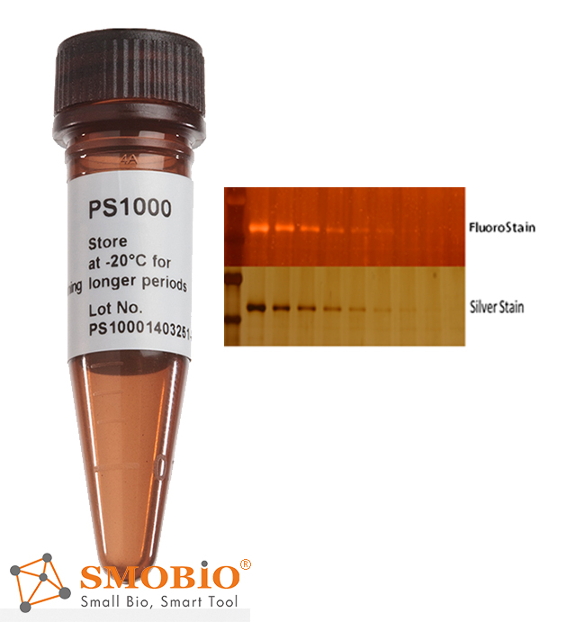
Storage
Protected from light -20°C for 24 months
Other Products
DNA Isothermal Rapid Amplification Kit (Colloidal Gold Test Strip Type)
Product Info
Document
Product Info
Product Details
Application:
DNA Nucleic Acid
Format:
NFO Kit
Kit Components:
Reagents,Buffer,Enzymes
Manufacturer:
Amp-future Bio
Product Name:
DNA Isothermal Rapid Amplification Kit (Colloidal Gold Test Strip Type)
Reaction Time:
5-20mins
Reaction Volume:
50μL
Sensitivity:
500-1000copies/ml
Shelf Life:
14 Months
Specificity:
High
Storage Conditions:
-20℃
Suitability:
Universal
High Light:
Isothermal DNA Amplification Kit
,
DNA Amplification Kit NFO
,
20mins nucleic acid amplification
Payment & Shipping Terms
Minimum Order Quantity
48T
Price
USD$3.8/T
Packaging Details
16T/Bag,48T/Box
Delivery Time
6 working days
Payment Terms
T/T, MoneyGram
Supply Ability
100000T/Month
Product Description
DNA Isothermal Rapid Amplification Kit (Colloidal Gold Test Strip Type)
【Principle Overview】
This kit is based on a rapid nucleic acid amplification technology at room temperature and constant temperature: at room temperature and constant temperature (generally 39℃~42℃), with the help of auxiliary proteins and single-strand binding proteins, the recombinase and primers form a complex; Source search and combine the target homology domain, at this time, a D-loop region is formed at the homology position and strand exchange begins; along with the dissociation of the recombinase from the complex, the polymerase also binds to the 3′ end of the primer and begins chain extension. Relying on the action of nfo enzyme, adding specific molecular probes designed according to the template, and using colloidal gold technology (sandwich method) can detect the final result.
【Primer design】
It is recommended to use primers with a length of 30-35 bp.Too short primers will affect the amplification speed and detection sensitivity;the 5′ end of the reverse primer is labeled with a modification group (commonly used biotin).
Primers are designed to avoid the formation of secondary structures that affect amplification; the length of the amplicon is recommended to be 150-500bp.
【Colloidal gold probe design】
In the middle of the forward and reverse primer,design a sequence of 46-52nt in length that is complementary to the target fragment; modify an antigen marker (typical FAM) at the 5′ end;mark a dSpacer (tetrahydrofuran, THF) in the middle of the 5′ end and the 3′ end ), as the recognition site of nfo;the 3′ end is labeled with a modification group, such as amine group, phosphate group or C3-Spacer, etc.
【Features】
This kit has the advantages of high sensitivity, strong specificity,and short reaction time (only 15 minutes),and the reaction components are in dry powder state,which is easy to operate and easy to store.
This reagent has low equipment requirement,and the reaction operation can be carried out in metal baths, water baths, etc.,and there is no need to purchase expensive exclusive equipment such as PCR amplifiers.
Composition
Content
A buffer
1.6mL×1Tube
B buffer
150μL×1Tube
Positive control template-II
100μL×1Tube
Positive control probe and primer mix-II
70μL×1Tube
Reagent
48 T
Guide Manual
1 Copy
【Kit storage】
1. Storage conditions: storage temperature ≤ -20℃constant temperature environment, keep away from light and avoid heavy pressure;
2. Product validity period: 14 months;
3. See the outer packaging for the production date.
Document
This kit is based on a rapid nucleic acid amplification technology at room temperature and constant temperature: at room temperature and constant temperature (generally 39℃~42℃), with the help of auxiliary proteins and single-strand binding proteins, the recombinase and primers form a complex; Source search and combine the target homology domain, at this time, a D-loop region is formed at the homology position and strand exchange begins; along with the dissociation of the recombinase from the complex, the polymerase also binds to the 3′ end of the primer and begins chain extension. Relying on the action of nfo enzyme, adding specific molecular probes designed according to the template, and using colloidal gold technology (sandwich method) can detect the final result.
Permagen’s 24 x 1.5 mL Microfuge Tube Magnetic rack is designed for magnetic bead separations from up to 24 tubes
Accommodates many common 1.5 mL Microcentrifuge and some 2.0 mL tubes
Tubes are angled and beads will be pulled to back wall allowing easy aspiration and tip tracking down the front wall of the tubes without disturbing bead pellet
Food and beverage samples such as milk or milk based products, wine, fruit juices, paper (and cardboard), waste treatment samples and other materials (e.g. biological cultures etc.).
This product has been discontinued.
The hydrogen peroxide Assay Kit provides a simple robust method for the measurement of hydrogen peroxide. This kit utilizes a highly sensitive Megaplex Red probe which in the presence of horseradish peroxidase (HRP), allows for the measurement of H2O2 in a sample. Upon oxidation of the probe by HRP, a coloured product (resorufin) is formed that can be measured in fluorescence mode using excitation in the range 530-560 nm and emission at ~ 590 nm or colourimetrically at 570 nm. The excitation and emission maxima of Megaplex Red are 571 nm and 585 nm respectively.
Fluorometric/UV method for the determination of H2O2 content in a variety of samples.
Advantages
Simple format
Very competitive price (cost per test)
Ability to run in fluorescence or absorbance mode
User friendly – Detailed protocol provided for the creation of calibration curve and the calculation of concentration in fluorescence mode. Single point standard for quicker and simpler analysis in absorbance mode.
Mega-Calc™ software tool is available from our website for hassle-free raw data processing
Suitable for manual, microplate and auto-analyser formats
All reagents stable for > 2 years
Document
The hydrogen peroxide Assay Kit provides a simple robust method for the measurement of hydrogen peroxide. This kit utilizes a highly sensitive Megaplex Red probe which in the presence of horseradish peroxidase (HRP), allows for the measurement of H2O2 in a sample. Upon oxidation of the probe by HRP, a coloured product (resorufin) is formed that can be measured in fluorescence mode using excitation in the range 530-560 nm and emission at ~ 590 nm or colourimetrically at 570 nm. The excitation and emission maxima of Megaplex Red are 571 nm and 585 nm respectively.