

Description
Q-PAGE™ Bis-Tris Precast Gel is a high-performance and easy to use precast polyacrylamide gel for electrophoresis in Bis-Tris buffer system (MOPS or MES). The optimized gel formula allows Q-PAGE™ Bis-Tris Precast Gel to show improved resolution, accurate results, and an extended shelf-life over conventional Laemmli Tris-HCl gels.
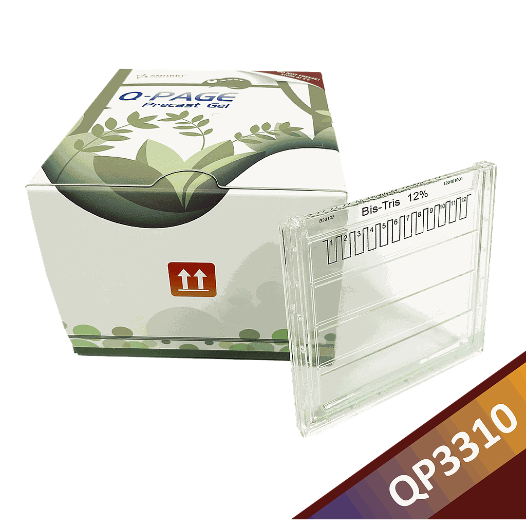
Q-PAGE™ Bis-Tris Precast Gels are available in gradient (4 to 12%) and fixed (8% and 12%) concentrations of polyacrylamide in 12-and 15-well formats. Two available cassette sizes, Mini (10 x 8.3 cm) and Midi (10 x 10 cm), are compatible with most popular protein electrophoresis systems. Q-PAGE™ Mini (QP2XXX) Gels are suitable for Bio-Rad® and other systems. Q-PAGE™ Midi (QP3XXX) Gels are suitable for Invitrogen® XCell SureLock® Mini-Cell, Invitrogen® Mini Gel Tank, Hoefer SE260, and other systems.
Key Features
- User-friendly gel cassette:
- Numbered and framed wells for sample loading
- With cassette opener for easy use
- Enhanced gel performance:
- Enhanced band sharpness
- Better resolution of small proteins
- Stable for shipping at ambient temperature
- Easy compatibility:
- Available as homogeneous and adjusted gradient gels for a wide range of protein separation.
- Compatible with most popular protein electrophoresis systems
Storage and stability
Store Q-PAGE™ Precast Gels at 4°C for periods up to 12 months.
Do not freeze Q-PAGE™ Precast Gels Remove tape and comb before electrophoresis.
Keep Q-PAGE™ Precast Gels flat during storage.


