

Payment & Shipping Terms
Minimum Order Quantity
48T
Price
USD$3.8/T
Packaging Details
16T/Bag,48T/Box
Delivery Time
6 working days
Payment Terms
T/T, MoneyGram
Supply Ability
100000T/Month
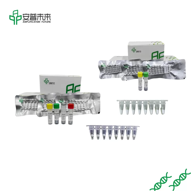
Product Description
Amplification kits come in a variety of reagent forms such as freeze-dried powder, freeze-dried microspheres, and master mix. (This paragraph can be marked in different forms)
product:
DNA Isothermal Rapid Amplification Kit (Fluorescent type)
DNA Isothermal Rapid Amplification Kit (Colloidal gold test strip type)
RNA Isothermal Rapid Amplification Kit (Basic Type)-II
…
Nucleic acid detection process: sampling, nucleic acid extraction, nucleic acid amplification, and result analysis. There are corresponding products that can be applied for the experimental steps after the sampling operation, which are more suitable for on-site rapid inspection needs.


