


DD5 Low Speed 5000rpm Swing rotor 4x800ml lab Centrifuge Adapters for 5/10/15/50ml tubes.

DD5 Low Speed Large Capacity Centrifuge Application:
DD5 Lab centrifuge widely used in the fields of pharmaceutical factory and laboratory, can hold swing out rotors and angle rotors both.
DD5 Low Speed Large Capacity Centrifuge Features:
1. Brushless frequency motor which in great torque, free maintenance, no powder pollution, quick in speed up and down.
2. Digital display which indicates the program, speed, time, RCF and temperature.
3. Micro computer control, there are 10 kinds of program and 10 kinds of acceleration/deceleration for your choice.
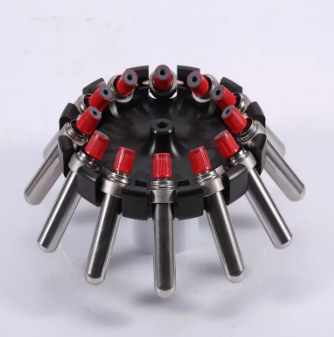
4. There are kinds of rotors for your choice.
5. Electric lid lock, compact design, super speed and imbalance protection.
6. The centrifuge body is made of high-quality steel, safe and reliable.
DD5 Low Speed Laboratory Centrifuge Technical Parameters:
| Max. Speed | 5000rpm |
| Max.RCF | 4730xg |
| Max.Capacity | 4x800ml |
| Timer | 0~99mins |
| RPM/RCF Convert | Yes |
| Noise(DB) | ≤58 |
| Acc/Dec | 10 Rates |
| Speed Accuracy | ±20rpm |
| Voltage(V/HZ) | AC 220V/110V 50HZ/60HZ |
| Dimension(LxWxH mm) | 720x540x830 |
| Net Weight(Kg) | 108kg |
| Certificates | CE,ISO |
| Warranty | 1 Year |
DD5 Low Speed Laboratory Centrifuge Matched Rotors:
| Order No. | Rotor type | Max.Speed (rpm) | Max.Volume (ml) | Max.RCF (xg) |
| No30671 | Swing out rotor | 4,000 | 4x800ml(round cup) | 3,450 |
| No30679 | Swing out rotor | 4,000 | 4x500ml(square cup) | 3,450 |
| No30696 | Swing out rotor | 4,000 | 4x500ml(round cup) | 3,530 |
| No31377 | Swing out rotor | 5,000 | 4x50ml | 4,730 |
| 4x100ml | 4,730 | |||
| No30592 | Swing out rotor | 4,000 | 4x30x7ml | 3,140 |
| 4x40x7ml | 3,270 | |||
| 4x18x10ml | 3,140 | |||
| No31491 | Mircoplater rotor | 4,000 | 2x4x96well | 2,910 |
| No31494 | Microplater rotor | 4,000 | 4x4x96well | 2,940 |
| No30627 | Angle rotor | 5,000 | 30x15ml | 3,830 |
| No30641 | Angle rotor | 5,000 | 12x50ml | 3,860 |
| No30642 | Angle rotor | 4,000 | 24x50ml | 2,970 |
| No30614 | Angle rotor | 5,000 | 6x100ml | 3,130 |
| No30643 | Angle rotor | 4,000 | 12x100ml | 2,970 |
The Corners of our Company____________________________________

A rapid, high performance dye-terminator removal process based on the paramagnetic bead technology. The paramagnetic bead format requires no centrifugation or filtration and is easily performedmanually or fully automated for high throughput dye-terminator removal. Compared to similar systems, this product produces sequences with longer Phred 20 read lengths and higher signal intensities than any other purification technology.
Specifications
| Features | Specifications |
| Main Functions | Removal of free fluorescent dye from sequencingsolution (Replace Beckmen or agencourt CleanSeq) |
| Applications | Automated sequences |
| Purification technology | Magnetic beads technology |
| Process method | Manual or automatic |
| Sample type | DNA doped with fluorescentdye |
| Sample amount | 5μl |
| Recovery | 90% |
| Elution volume | ≥25μl |
| Operation time | ≤50 minutes |
The CleanSeq method contains magnetic particles in an optimized binding buffer to selectively capture sequencing extension products. The protocol can be performed directly in the thermal cycling plate. Unincorporated dyes, nucleotides, salts and contaminants are removed using a simple washing procedure. The purification procedure is amenable to a variety of automation platforms since it requires no centrifugation or vacuum filtration.
Advantages
Kit Contents
| Contents | BCS-5 | BCS-50 | BCS-500 |
| CleanSeq Beads | 5 ml | 50 ml | 500 ml |
Storage and Stability
CleanSeq Beads should be stored at 2-8°C upon arrival and is stable up to 6 months under the condition. However, short-term storage (up to 12 weeks) at room temperature (15-25°C) does not affect its performance. Mix CleanSeq Beads well before using. The reagent should appear homogenous and consistent in color.
DO NOT FREEZE.
A rapid, high performance dye-terminator removal process based on the paramagnetic bead technology. The paramagnetic bead format requires no centrifugation or filtration and is easily performedmanually or fully automated for high throughput dye-terminator removal. Compared to similar systems, this product produces sequences with longer Phred 20 read lengths and higher signal intensities than any other purification technology.

| Clone | IHC411 |
| Source | Rabbit Monoclonal |
| Positive Control | Tonsil, Lung Adenocarcinoma |
| Dilution Range | 1:200 |
Programmed Death-Ligand 1 (PD-L1), also known as CD274 or B7 Homolog 1 (B7-H1), is a transmembrane protein involved in suppressing the immune system and rendering tumor cells resistant to CD8 T cell-mediated lysis through binding of the Programmed Death-1 (PD-1) receptor. Overexpression of PD-L1 may allow cancer cells to evade the actions of the host immune system. In renal cell carcinoma, upregulation of PD-L1 has been linked to increased tumor aggressiveness and risk of death, and, in ovarian cancer, higher expression of this protein has lead to significantly poorer prognosis. PD-L1 has also been linked to systemic lupus erythematosus and cutaneous melanoma. When considered in adjunct with CD8 tumor-infiltrating lymphocyte density, expression levels of PD-L1 may be a useful predictor of multiple cancer types, including stage III non-small cell lung cancer, hormone receptor negative breast cancer, and sentinel lymph node melanoma.