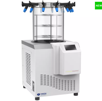
Extreme vacuum 4 * 10-4mbar,with a pumping capacity of 78L~200L/optional pump of various types
Vacuum pump connection pipe
Single forming stainless steel flexible pipe DN16 ISO-KF L-1.2m
anti-corrosive treatment
Cold trap, condensing coil/all equipped with PTFE anti-corrosion treatment as standard. Freeze-drying organic solvents
External valve
6/optional
1-48 pieces/optional
Eutectic point detection system
Support/Optional
Data output analysis
Support/Optional
Power
0.6kw
1.1kw
0.5kw
Other Products
[PM2610] ExcelBand™ Enhanced 3-color High Range Protein Marker (9-245 kDa), 250 μl x 2
Product Info
Document
Product Info
Description
The PM2610/PM2611 ExcelBand™ Enhanced 3-color High Range Protein Marker is a ready to use three-color protein standard with 12 pre-stained proteins covering a wide range of molecular weights from 10 to 245 kDa in Tris-Glycine buffer (9 to 235 kDa in BisTris (MOPS) buffer and 10-235 kDa in Bis-Tris (MES) buffer). Proteins are covalently coupled with different chromophores for easy identification of bands, with two reference proteins carrying enhanced intensity corresponding to a green at 25 kDa and red at 75 kDa, respectively, as separated on SDS-PAGE (Tris-Glycine buffer). The ExcelBand™ Enhanced 3-color High Range Protein Marker is designed for monitoring protein separation during SDS-polyacrylamide gel electrophoresis, verification of Western transfer efficiency on membranes (PVDF, nylon, or nitrocellulose) and for approximating the size of proteins.
Features
Ready-to-use — Premixed with a loading buffer for direct loading, no need to boil.
Two reference bands — 75 kDa (red) and 25 kDa (green)
Contents
Approximately 0.2~0.6 mg/ml of each protein in the buffer (20 mM Tris-phosphate (pH 7.5 at 25°C), 2 % SDS, 3.6 M Urea, and 15 % (v/v) Glycerol).
Quality Control
Under suggested conditions, the PM2610/PM2611 Enhanced 3-color High Range Protein Marker resolves 12 major bands in 15% SDS-PAGE (Tris-Glycine buffer, MOPS, and MES buffer) and after Western blotting to nitrocellulose membrane.
Storage
4°C for 3 months -20°C for long term storage
Document
The PM2610/PM2611 ExcelBand™ Enhanced 3-color High Range Protein Marker is a ready to use three-color protein standard with 12 pre-stained proteins covering a wide range of molecular weights from 10 to 245 kDa in Tris-Glycine buffer (9 to 235 kDa in BisTris (MOPS) buffer and 10-235 kDa in Bis-Tris (MES) buffer). Proteins are covalently coupled with different chromophores for easy identification of bands, with two reference proteins carrying enhanced intensity corresponding to a green at 25 kDa and red at 75 kDa, respectively, as separated on SDS-PAGE (Tris-Glycine buffer). The ExcelBand™ Enhanced 3-color High Range Protein Marker is designed for monitoring protein separation during SDS-polyacrylamide gel electrophoresis, verification of Western transfer efficiency on membranes (PVDF, nylon, or nitrocellulose) and for approximating the size of proteins.
Compatible with mass spectrometry and NMR spectroscopy
Purification is based on spin column chromatography that uses Norgen’s proprietary resin separation matrix
This kit provides a fast and simple procedure for the isolation of total proteins from tissue, bacteria, yeast or mammalian cells without the use of SDS, Triton® X-100 and other detergents. Detergents are extensively used to prepare protein samples; however, these detergents have undesirable effects on downstream analysis. These effects include extraneous peaks in mass spectrometry, artifacts with chromatography and electrophoresis, interference with microinjection into cells and interference with protein immunization.
The Detergent-Free Total Protein Isolation Kit maintains high protein recovery and yields proteins that are 100% detergent-free. Purification is based on using Norgen’s proprietary resin together with Lysis Solution, followed by protein filtration using a filter column (provided). The purified proteins can be used in a number of downstream applications including mass spectrometry, SDS-PAGE, isoelectric focusing, NMR spectroscopy and more.
Each Lysis Tube is able to process up to 50 mg of tissue, 1010 bacterial cells, 109 yeast cells or 107 mammalian cells. Preparation time for 12 samples is less than 10 minutes.
Maximum Amount Of Starting Material: Tissues Animal Cells Bacteria Yeast
50 mg 1 x 10⁷ cells 1 x 10¹⁰ cells 1 x 10⁹ cells
Time to Process 12 Samples
Less than 10 minutes
Storage Conditions and Product Stability The Lysis Solution should be kept tightly sealed and stored at room temperature. Once opened, the solution should be stored at 4°C. This kit is stable for 2 years after the date of shipment.
Blood samples contain rich DNA, including mitochondrial DNA, genomic DNA, circulating DNA (mostly released into blood after tumor cell apoptosis) in white blood cells, as well as parasitic viral or microbial DNA. These DNA are important parameters in clinical testing or diagnosis, which are also valuable materials for medical research. There are three main issues with extracting DNA from blood samples:
1. The sample is highly infectious, posing great harm to operators and the environment.
2. The source of DNA is complex and aportion of the nucleic acid, such as viral DNA or free DNA, may be lost during the operation, leading to downstream detection failure;
3. Blood sample contains a large amount of impurities and inhibitory factors.
Currently there are many methods available for extracting DNA from whole blood samples, such as phenol chloroform extraction, salting out method, etc. However, these methods require pre-treatment of blood sample, which removes red blood cells and isolate white blood cells in the first step. Due to the requirement that it cannot inactivate or kill pathogens during the process of removing red blood cells, the waste liquid (red blood cell lysate) and consumables may be contaminated by pathogens and become infectious, posing a danger to the entire laboratory environment and operators. In addition, during the process of removing red blood cells, useful nucleic acid information such as viruses, microorganisms, or circulating DNA is also lost, leading to experiment or detection failures.
The HiPure Blood DNA Kits series provided by Magen Company uses silica gel column purification technology, which can directly lyse whole blood samples without the need for white blood cell separation. Whole blood samples are directly mixed with lysates and proteases, resulting in the inactivation of pathogens, greatly reducing the infectivity, environmental pollution, and the chance of operators being infected. Due to the direct lysis and digestion of samples, except lymphocyte DNA, other circulating DNA as well as DNA from viruses and microorganisms, can also be recovered.
This product provides fast and easy methods for purification of total DNA for reliable PCR and Southern blotting. Total DNA (e.g., genomic, viral, mitochondrial) can be purified from whole blood, tissue and culture cells.
Details
Specifications
Features
Specifications
Main Functions
Isolation total DNA from 10ml blood and 1g tissue using Maxi column
Applications
PCR, southern bolt and virus detection, etc
Purification method
Maxi spin column
Purification technology
Silica technology
Process method
Manual (centrifugation or vacuum)
Sample type
Tissue, cell, blood, saliva, swab, blood spot, semen and other clinical samples
Sample amount
3-10ml
Elution volume
≥700μl
Time per run
≤90 minutes
Liquid carrying volume per column
4ml
Binding yield of column
5mg
Principle
This product is based on silica column purification. The sample is lysed and digested with lysate and protease, DNA is released into the lysate. Transfer to an adsorption column. Nucleic acid is adsorbed on the membrane, while protein is not adsorbed and is removed with filtration. After washing proteins and other impurities, Nucleic acid was finally eluted with low-salt buffer (10mm Tris, pH9.0, 0.5mm EDTA).
Advantages
High quality DNA – meet a variety of downstream applications, including PCR, qPCR, enzyme digestion, hybridization, etc.
Fast – without separation of leukocytes, organic extraction or ethanol precipitation
Simple – all nucleic acids can be obtained by direct digestion
Pertinence – specially designed for isolating DNA from 3-10ml blood and related body fluids
Wide applicability – handle a variety of liquid samples
Proteinase K, RNase A should be stored at 2-8°C upon arrival. However, short-term storage (up to 12 weeks) at room temperature (15-25°C) does not affect their performance. The remaining kit components can be stored at room temperature (15-25°C) and are stable for at least 18 months under these conditions.
*Note:Leukocyte protocol can be used when large volume whole blood samples need to be processed. Whole blood was treated with red blood cell lysate, and white blood cells were obtained by centrifugation before extraction
Document
Blood samples contain rich DNA, including mitochondrial DNA, genomic DNA, circulating DNA (mostly released into blood after tumor cell apoptosis) in white blood cells, as well as parasitic viral or microbial DNA. These DNA are important parameters in clinical testing or diagnosis, which are also valuable materials for medical research. There are three main issues with extracting DNA from blood samples: