
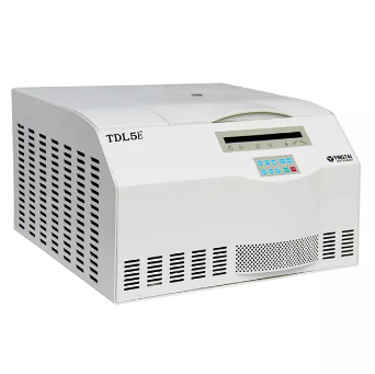

TDL5E Bench top low speed refrigerated centrifuge machine
Application:
Widely used in the field of biochemistry, biological products, pharmaceutical factory and laboratory.
TDL5E Technical Parameters:
| Max. Speed | 5000rpm | Max.RCF | 4420xg |
| Max. Volume | 4x180ml | Speed Accuracy | ±20rpm |
| Timer | 1~99h59min | Noise | ≤60dBA |
| Dimension(LxWxHmm) | 570x600x340mm | Net Weight | 76Kg |
| Power Supply | AC220V/110V 50HZ/60HZ | Temperature Range | -20℃~40℃ |
| Temperature Accuracy | ±1℃ | Certifications | CE, ISO & Calibration report is available. |
Matched Rotors for TDL5E:
| Order No. | Rotor Type | Max. Speed(rpm) | Volume(ml) | Max.RCF(xg) |
| 5E-1 | Swing rotor | 4000 | 4x180ml | 2580 |
| 5E-2 | Microplate rotor | 4000 | 2x3x96well | 2020 |
| 5E-3 | Swing rotor | 5000 | 4x1x50ml | 4420 |
| 5E-4 | Swing rotor | 5000 | 4x1x100ml | 4420 |
| 5E-5 | Swing rotor | 4000 | 4x2x50ml | 2820 |
| 5E-6 | Swing rotor | 4000 | 4x4x15ml | 2820 |
| 5E-7 | Swing rotor | 4000 | 4x6x10/7ml | 2580 |
| 5E-8 | Swing rotor | 4000 | 4x6x5ml | 2170 |
| 5E-9 | Swing rotor | 4000 | 4x4x10/7ml | 2580 |
| 5E-10 | Swing rotor | 4000 | 4x4x5ml | 2170 |
| 5E-11 | Fixed rotor | 5000 | 6x15ml | 2540 |
| 5E-12 | Fixed rotor | 5000 | 12x15ml | 3080 |
| 5E-13 | Fixed rotor | 5000 | 24x15ml | 3500 |
| 5E-14 | Fixed rotor | 5000 | 4x50ml | 2520 |
| 5E-15 | Fixed rotor | 5000 | 6x50ml | 2850 |
| 5E-16 | Fixed rotor | 5000 | 4x100ml | 2630 |
| 5E-17 | Fixed rotor | 5000 | 6x100ml | 3130 |


