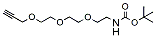
t-Boc-N-Amido-PEG3-propargyl enables formation of triazole linkage with azide-bearing compound via copper catalyzed Click Chemistry. Under mild acidic conditions, t-Boc group can be removed to yield the free amine. Reagent grade, for research purpose. Please contact us for GMP-grade inquiries.
Detail
t-Boc-N-Amido-PEG3-propargyl enables formation of triazole linkage with azide-bearing compound via copper catalyzed Click Chemistry. Under mild acidic conditions, t-Boc group can be removed to yield the free amine. Reagent grade, for research purpose. Please contact us for GMP-grade inquiries.
Other Products
[PM2500] ExcelBand™ 3-color Regular Range Protein Marker (9-180 kDa), 250 μl x 2
Product Info
Document
Product Info
Description
The PM2500 ExcelBand™ 3-color Regular Range Protein Marker is a ready-to-use three-color protein standard with 10 pre-stained proteins covering a wide range of molecular weights from 10 to 180 kDa in Tris-Glycine Buffer (9 to 170 kDa in Bis-Tris (MOPS) buffer and 10 to 170 kDa Bis-Tris (MES) buffer). Proteins are covalently coupled with a blue chromophore except for two reference bands (one green and one red band at 25 kDa and 75 kDa respectively) when separated on SDS-PAGE (Tris-Glycine buffer). PM2500 ExcelBand™ 3-color Regular Range Protein Marker is designed for monitoring protein separation during SDS-polyacrylamide gel electrophoresis, verification of Western transfer efficiency on membranes (PVDF, nylon, or nitrocellulose) and for approximating the size of proteins.
Features
Ready-to-use — Premixed with a loading buffer for direct loading, no need to boil.
Two reference bands — 75 kDa (red) and 25 kDa (green)
Contents
Approximately 0.1~0.4 mg/ml of each protein in the buffer (20 mM Tris-phosphate (pH 7.5 at 25°C), 2% SDS, 0.2 mM DTT, 3.6 M urea, and 15% (v/v) glycerol).
Quality Control
Under suggested conditions, PM2500 ExcelBand™ 3-color Regular Range Protein Marker resolves 10 major bands in 15% SDS-PAGE (Tris-Glycine buffer, MOPS, and MES buffer) and after Western blotting to nitrocellulose membrane.
Storage
4°C for 3 months -20°C for long term storage
Document
The PM2500 ExcelBand™ 3-color Regular Range Protein Marker is a ready-to-use three-color protein standard with 10 pre-stained proteins covering a wide range of molecular weights from 10 to 180 kDa in Tris-Glycine Buffer (9 to 170 kDa in Bis-Tris (MOPS) buffer and 10 to 170 kDa Bis-Tris (MES) buffer). Proteins are covalently coupled with a blue chromophore except for two reference bands (one green and one red band at 25 kDa and 75 kDa respectively) when separated on SDS-PAGE (Tris-Glycine buffer). PM2500 ExcelBand™ 3-color Regular Range Protein Marker is designed for monitoring protein separation during SDS-polyacrylamide gel electrophoresis, verification of Western transfer efficiency on membranes (PVDF, nylon, or nitrocellulose) and for approximating the size of proteins.
Nucleic acid testing (NAT) is the method of choice for detection and quantification of a wide range of micro organisms. Primerdesign manufactures and supplies high quality quantitative real-time PCR kits for the detection and simultaneous quantification of numerous significant pathogens . A copy number standard curve is provided for quantification and an the internal extraction template (DNA or RNA), controls for the quality of the nucleic acid extraction and eliminates false negative results.
The kit is designed with the broadest possible detection profile to ensure that all clinically relevant strains and subtypes are detected. Target sequences are selected by working with data from key opinion leaders in the field. Multiple sequence alignments and unprecedented real-time PCR expertise in design and validation ensure the best possible kit. Details of the target and priming specificity are included in the individual handbooks above.
Packaged, optimised and ready to use. Expect Better Data.
Document
Exceptional value for money Rapid detection of all clinically relevant subtypes Positive copy number standard curve for quantification Highly specific detection profile High priming efficiency Broad dynamic detection range (>6 logs) Sensitive to < 100 copies of target
Accurate controls to confirm findings
150 reactions
Dietary fiber can generally be described as the carbohydrate content of food that is not digested in the human small intestine. It passes into the large intestine where it is partially or fully fermented. These characteristics of dietary fiber are associated with its numerous well documented health benefits.
Dietary Fiber is a mixture of complex organic substances, including hydrophilic compounds, such as soluble and insoluble polysaccharides and non-digestable oligosaccharides, as well as a range of non-swellable, more or less hydrophobic, compounds such as cutins, suberins and lignins. The procedures for the determination and analysis of total dietary fiber as outlined in our assay protocol are based on the methods of Lee et al.1 and Prosky et al.2,3 (AOAC 991.43, AOAC 985.29, AACC 32-07.01 and AACC 32-05.01). However, the enzymes in the Megazyme Total Dietary Fiber Kit can also be used in other dietary fiber analytical methods such as AACC Method 32-21.01 and AACC Method 32-06.01.
1. Association of Official Analytical Chemists. (1985). Official Methods of Analysis, 14th ed., 1st suppl. Secs. 43, A14-43, A20, p.399. 2. Association of Official Analytical Chemists. (1986). Changes in methods. J. Assoc. Off. Anal. Chem., 69, 370. 3. Association of Official Analytical Chemists. (1987). Changes in methods. J. Assoc. Off. Anal. Chem., 70, 393.
See General Referee Reports: Journal of AOAC INTERNATIONAL, Vol. 81, No. 1, 1998.
Two separate methods are described in the associated assay protocol:
METHOD 1: DETERMINATION OF TOTAL, SOLUBLE AND INSOLUBLE DIETARY FIBER Based on AOAC Method 991.43 “Total, Soluble, and Insoluble Dietary Fiber in Foods” (First Action 1991) and AACC Method 32-07.01 “Determination of Soluble, Insoluble, and Total Dietary Fiber in Foods and Food Products” (Final Approval 10-16-91).
METHOD 2: DETERMINATION OF TOTAL DIETARY FIBER Based on AACC method 32-05.01 and AOAC Method 985.29.
Note that a letter of endorsement from the original method developer, Dr. Leon Prosky, is included in the Documents Tab.