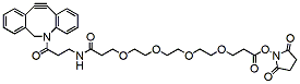
DBCO-NHCO-PEG4-NHS ester is a PEG linker containing NHS ester that is able to react specifically and efficiently with primary amines such as the side chain of lysine residues or aminosilane-coated surfaces at neutral or slightly basic condition to form a covalent bond. The hydrophilic PEG spacer arm improves water solubility and can also provide a long and flexible connection that minimizes steric hindrance during ligation. DBCO is commonly used for copper-free Click Chemistry reactions. Reagent grade, for research purpose. Please contact us for GMP-grade inquiries.
Detail
DBCO-NHCO-PEG4-NHS ester is a PEG linker containing NHS ester that is able to react specifically and efficiently with primary amines such as the side chain of lysine residues or aminosilane-coated surfaces at neutral or slightly basic condition to form a covalent bond. The hydrophilic PEG spacer arm improves water solubility and can also provide a long and flexible connection that minimizes steric hindrance during ligation. DBCO is commonly used for copper-free Click Chemistry reactions. Reagent grade, for research purpose. Please contact us for GMP-grade inquiries.
Other Products
CRM003 E. coli and Coliform Chromogenic Agar
Product Info
Document
Product Info
Introduction
Usages:
For the rapid detection and of coliforms and E. coli.
Principle:
Peptone and yeast extract powder provides carbon and nitrogen sources and trace elements; sodium chloride maintains osmotic equilibrium; agar as medium coagulant; dodecyl sulfate inhibit Gram-positive bacteria; chromogenic substrate were mixed occurrence of coliforms and E. coli enzyme corresponding specific reactions, hydrolysis of the substrate, the release of the color groups, in a pale yellow plates coliforms appears orange-red colonies while E.coli appears blue-green colonies.
Formulation (per liter):
Peptone 15.0g
Yeast extract powder 3.0g
Sodium chloride: 5.0g
Sodium lauryl sulfate: 0.1g
Agar: 12.0g
Mixed chromogenic substrate: 6.77g
Final pH 7.0 ± 0.2
How to use: 1. Weigh 41.9g of the product, adding , 1.0 L of distilled or deionized water, heated to boiling stir until completely dissolved, dispensing into flask, 115 autoclaved 10minutes.
2, Take 25.0g or 25.0mL of sample with sterile procedures, added to the flask containing 225.0mL of sterile phosphate buffered saline (or saline) ,shaken thoroughly homogenized with a homogenizer or a 1:10 dilution of 1min solution, diluted 1:10 and then continue to select the appropriate serial dilutions of three, the two plates inoculated with each dilution, poured dissolved by heating and cooled to about 45 medium.
3, observe the results.
Quality control:
This product appears light yellow after pouring on plate, these strains were inoculated after 36 ± 1 18 ~ 24h culture growth in the following table.
Bacteria name bacteria NO. growth situation feature
Escherichia coli ATCC25922 good blue-green colonies
Citrobacter ATCC8090 good orange-red colonies
Salmonella typhimurium CMCC50115 good colorless colonies
Enterococcus faecalis ATCC29212 suppressed —–
Storage: Store in a dark, cool and dry place, tighten the caps immediately after use. Storage period of two years.
Salmonella spp. are members of the family Enterobacteriaceae. They are Gram-negative, facultatively anaerobic, flagellated, rod-shaped organisms. They are approximately 0.7 to 1.5 µm in diameter and 2 to 5 µm in length and responsible for a large number of cases of foodborne illness throughout the world. Salmonella have circular DNA genomes with a mean length of approximately 4530 kb, although this can vary by up 1000 kb. Salmonella classification is extremely complex, however, the genus is divided into two species: S. enterica and S.bongori. S. enterica is then itself divided into 6 biochemically distinct subspecies and the Salmonella genus is further classified into serovars (serotypes) based on the lipopolysaccharide (O), flagella protein (H), and sometimes the capsular (VI) antigens. There are more than 2500 known serovars and within a serovar there may be strains that differ in virulence.
Salmonella are mainly transmitted by the faecal-oral route. They are carried asymptomatically in the intestines or gall bladder of many animals, being continuously or intermittently shed in the faeces. Humans can become infected if they do not wash their hands after contact with infected animals or animal faeces. In such instances the bacteria adhere to and enter the cells of the intestinal epithelium. The toxins produced by the bacteria can damage and kill the cells that line the intestines, which results in intestinal fluid loss. The bacteria can survive for weeks in a dry environment and far longer in water thus they are frequently present in polluted waters. Salmonella can also be carried latently in the mesenteric lymph nodes or tonsils; these bacteria are not shed, but can become reactivated after stress or immunosuppression. In addition, fomites and vectors can spread Salmonella and vertical transmission occurs in birds, with contamination of the vitalize membrane, albumen and possibly the yolk of eggs. Salmonella spp. can also be transmitted in utero in mammals.
There are two different disease conditions that are distinct to salmonellosis; gastroenteritis and enteric typhoid fever. The gastroenteritis is a nonsystemic infection of the intestinal tract and regional lymph nodes that gives rise to headache, muscle aches, diarrhoea, vomiting, abdominal cramping, chills, fever, nausea and dehydration. In contrast, the enteric typhoid fever is a systemic disease in which the microorganism replicates within the cells of the reticuloendothelial system. The symptoms usually appear 6 to 72 hours after ingesting contaminated food although individuals can be infected with the bacteria without having symptoms. Those with and without symptoms shed the bacteria in their stool and it is important that personal hygiene be maintained at all times.
Document
Exceptional value for money Rapid detection of all clinically relevant subtypes Positive copy number standard curve for quantification Highly specific detection profile High priming efficiency Broad dynamic detection range (>6 logs) Sensitive to < 100 copies of target
Accurate controls to confirm findings
Ensure optimal results for sensitive applications like NGS
Up to a tenfold increase in microRNA mapping during sequencing runs to reduce costs
Isolates all sizes of exosomal and extracellular vesicle RNA, including microRNA.
Bind and elute all RNA irrespective of size or GC content, without bias.
No phenol extractions.
No Proteinase K treatment.
No carrier RNA.
Concentrate isolated RNA into a flexible elution volume ranging from 50 μL to 100 μL.
Purify high-quality RNA in 15-20 minutes.
Purified RNA is suitable for a variety of downstream applications, including Small RNA Sequencing.
Purification is based on EXTRAClean spin column chromatography that uses Norgen’s proprietary separation matrix.
The kit is designed to isolate all sizes of RNA, including microRNA. The kit provides a clear advantage over other available kits in that it does not require any special instrumentation, protein precipitation reagents, extension tubes, phenol/chloroform or protease treatments. Moreover, the kit allows the user to elute into a flexible elution volume ranging from 50 μL to 100 μL. The purified RNA is of the highest integrity, and can be used in a number of downstream applications including real time PCR, reverse transcription PCR, Northern blotting, RNase protection and primer extension, and expression array assays.
Exosomes purified using Norgen’s Purification Kits
Size of RNA Purified
All sizes, including miRNA and small RNA (< 200 nt)
Elution Volume
50-100 μL
Time to Complete 10 Purifications
35-40 minutes
Average Yields
Variable depending on specimen
Storage Conditions and Product Stability All buffers should be kept tightly sealed and stored at room temperature. This kit is stable for 2 years after the date of shipment. It is recommended to warm Lysis Buffer A for 20 minutes at 60°C if any salt precipitation is observed.