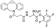
Sulfo DBCO-TFP Ester is a water-soluble, amine-reactive labeling reagent that enables simple and efficient incorporation of Sulfo DBCO moiety onto amine-containing molecules. The hydrophilic, sulfonated spacer arm greatly improves water solubility of DBCO derivatized molecules, in many cases making them completely soluble in aqueous media. A short spacer arm adds minimal mass to modified molecules.
Detail
Sulfo DBCO-TFP Ester is a water-soluble, amine-reactive labeling reagent that enables simple and efficient incorporation of Sulfo DBCO moiety onto amine-containing molecules. The hydrophilic, sulfonated spacer arm greatly improves water solubility of DBCO derivatized molecules, in many cases making them completely soluble in aqueous media. A short spacer arm adds minimal mass to modified molecules.
The Opentrons Flex 96-Channel pipette enables accurate high throughput pipetting, optimized for working in 96-well plates. Flex Pipettes use air displacement technology to offer highly accurate pipetting with pipette volume ranges from 5 – 1000 μL.
Smart sensors support automatic calibration, real-time positioning, and error detection. The 96-channel pipette occupies both pipette mounts and can easily be removed and swapped to an alternate pipette configurations to adapt your workflow needs.
Four 96-Channel Tip Rack Adapters are included with the purchase of one Flex 96-Channel Pipette.
Document
The Opentrons Flex 96-Channel pipette enables accurate high throughput pipetting, optimized for working in 96-well plates. Flex Pipettes use air displacement technology to offer highly accurate pipetting with pipette volume ranges from 5 – 1000 μL.
Smart sensors support automatic calibration, real-time positioning, and error detection. The 96-channel pipette occupies both pipette mounts and can easily be removed and swapped to an alternate pipette configurations to adapt your workflow needs.
Four 96-Channel Tip Rack Adapters are included with the purchase of one Flex 96-Channel Pipette.
Room Temperature DNA Amplification Kit For Isothermal Nucleic Acid
Product Info
Document
Product Info
Product Description
Room Temperature DNA Amplification Kit for Isothermal Nucleic Acid
Product Detail
Kit Storage and term of Validity
Storage term: stored at ≤-20℃,keep away from light, avoid heavy weight and repeated freezing and thawing.
Term of Validity: 14 months
Isothermal nucleic acid Principle Summary
The kit is based on room and constant temperature nucleic acid rapid amplification technology, its principle is that at room and constant temperature, the recombinase and primer form a protein/single-stranded nucleotide complex Rec/ssDNA, and invade the double-stranded DNA template with the help of auxiliary proteins and single-stranded binding protein SSB; then form a D-loop region at the invasion point and start to scan the DNA duplex, after finding the target region complementary to the primer and disintegration of the complex Rec/ssDNA, the polymerase also binds to the 3′ end of the primer to start the chain extension. The kit relies on the role of NFO enzyme and adds the designed specific molecular probes according to the template, and get the result by colloidal gold technology (sandwich method).
Isothermal nucleic acid Product Features
1/ High sensitivity and specificity, short reaction time.
2/ The reagent form is freeze-dried, stable and easy to operate.
3/ The reaction can be operated by metal bath and water bath pot without purchasing expensive PCR apparatus.
Technical Parameters:
Parameters
Details
Product Name
DNA Isothermal Amplification Kit NFO
Manufacturer
Amp-future
Storage Temperature
-20°C
Kit Components
Enzymes, Buffers ,Reagents
Packaging
48 Tests/box
Detection Limit
500-1000copies/µL
Shipping
ICE
Test Time
5-20mins
Isothermal nucleic acid Applications
Suitable for DNA isothermal rapid amplification kit(NFO type)
Primer: Require pair of nucleotide primers with the length of 25-35 bp.
DNA basic kit reaction temperature is 39 to 42℃ and time is 5-20 minutes.
Notes
1/ Please avoid nucleic acid contamination and set blank control during reaction due to the high sensitivity of the kit.
2/ Please take out the required quantity of MIRA reaction units for the experiment, and put the rest under storage conditions when performing the experiment.