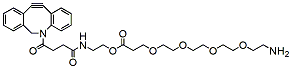
DBCO-C2-PEG4-amine is a PEG linker which contains DBCO and amine moieties. The DBCO group is commonly used in copper-free Click Chemistry reactions. The amine group is reactive with carboxylic acids, activated NHS esters, carbonyls (ketone, aldehyde) etc. The hydrophilic PEG spacer increases solubility in aqueous media. Reagent grade, for research purpose. Please contact us for GMP-grade inquiries.
Detail
DBCO-C2-PEG4-amine is a PEG linker which contains DBCO and amine moieties. The DBCO group is commonly used in copper-free Click Chemistry reactions. The amine group is reactive with carboxylic acids, activated NHS esters, carbonyls (ketone, aldehyde) etc. The hydrophilic PEG spacer increases solubility in aqueous media. Reagent grade, for research purpose. Please contact us for GMP-grade inquiries.
Other Products
16S V1-V3 Library Preparation Kit for Illumina
Product Info
Document
Product Info
Overview
Protocol optimized for DNA isolated from a diversity of samples including stool, soil, water, saliva, plant, urine, skin, and more
Simple and quick workflow: library could be prepared in less than 5 hours
Component of Norgen’s metagenomics workflow
A single NGS run can be prepared with up to 384 unique dual-index libraries
The 16S V1-V3 Library Preparation Kit for Illumina consists of the reagents and components required for library preparation of the 16S V1-V3 amplicon libraries to be used for next-generation sequencing on Illumina platforms. All molecular reagents including primers, enzyme mixes, indexes, and buffers are provided. Instructions for PCR clean up with the AMPure XP Magnetic Beads (supplied by customer) are also included for rapid purification of nucleic acid products generated at two steps of the workflow. The library prep workflow could be used for purified DNA inputs from different sources including stool, soil, water, saliva, plant, urine, skin swab, vaginal swab, cheek swab, nasal swab, plasma/serum, tongue swab, gum swab, and others.
The 16S V1-V3 Library Preparation Kit for Illumina has a streamlined procedure that reduces the handling time such that the library prep procedure can be completed in approximately 4 hours (see diagram below). Input DNA is first subjected to targeted PCR to amplify the V1-V3 region of the DNA encoding 16S rRNA. The post-PCR reaction is then cleaned up using AMPure XP beads. Dual index primers are then added using a limited-cycle PCR. The indexed amplicons flanked by 5′ and 3′ barcoded adaptors are then cleaned using AMPure XP beads. The libraries are then ready for quantification, pooling and sequencing.
Storage Conditions and Product Stability Norgen’s 16S V1-V3 Library Prep Kit for Illumina is shipped as one kit box (for the 24 prep kit) or two sub-component kits (for the 96 prep kit). All kits should be stored at -20°C upon arrival.
All kit components should remain stable for at least 1 year when stored at the specified storage conditions.
High yield and high quality genomic DNA with no RNA or protein contamination
DNA ready for any application including PCR, qPCR, genotyping and more
Norgen’s Cells and Tissue DNA Isolation Kits are designed for the rapid preparation of genomic DNA from cultured cells as well as various tissue samples and urine. The purified genomic DNA is fully digestible with all restriction enzymes tested, and is completely compatible with PCR and Southern Blot analysis.
Cells and Tissue DNA Isolation Kit (Spin Column)
Purification is based on spin column chromatography as the separation matrix. Norgen’s columns bind DNA under optimized salt concentrations and release the bound DNA under low salt and slightly alkali conditions. The protocol can be completed in approximately 30 minutes for cells and within 90 minutes for tissues. Each kit contains sufficient materials for 50 preparations.
Cells and Tissue DNA Isolation Micro Kit (Micro)
Optimized for small inputs of cells and tissues, such as Laser-Captured Microdissection (LCM). Purification is based on spin column chromatography as the separation matrix. Norgen’s columns bind DNA under optimized salt concentrations and release the bound DNA under low salt and slightly alkali conditions. Preparation time for a single sample is approximately 60 minutes, and each kit contains sufficient materials for 50 preparations.
Cells and Tissue DNA Isolation Kit (Magnetic Bead System)
Purification is based on the use of magnetic beads that bind DNA under optimized binding conditions. Norgen’s Cells and Tissue DNA Isolation Kit (Magnetic Bead System) allows for the isolation of genomic DNA from various types of animal tissues or cell samples. Preparation for 10 purifications is approximately 40 minutes of hands-on time.
Cells and Tissue DNA Isolation 96-Well Kit (High Throughput Magnetic Bead System)
Purification is based on the use of magnetic beads that bind DNA under optimized binding conditions. Norgen’s Cells and Tissue DNA Isolation 96-Well Kit (Magnetic Bead System) allows for the isolation of genomic DNA from various types of animal tissues or cell samples. The Cells and Tissue DNA Isolation 96-Well Kit (Magnetic Bead System) also can be integrated with a robotic automation system.
20 mg of animal tissue Up to 150 μL of viral suspension 3 x 10⁶ cells
Column Binding Capacity
> 50 μg
Average Yield
8 μg (from 1 x 10⁶ HeLa Cells) 10 μg (from 10 mg kidney)
Elution Volume
50 – 200 μL
Analyte Purified
Genomic DNA, mitochondrial DNA, viral DNA
Format
Spin Column
Time to Complete 10 Purifications
30 min (cells) and 90 min (tissue)
Storage Conditions and Product Stability All solutions should be kept tightly sealed and stored at room temperature. This kit is stable for 1 year after the date of shipment. The kit contains a ready-to-use Proteinase K, which is dissolved in a specially prepared storage buffer. The buffered Proteinase K is stable for up to 1 year after the date of shipment when stored at room temperature.
Propargyl-PEG14-t-butyl ester consists of a propargyl group and a t-butyl protected carboxyl group. The propargyl group can be used in copper catalyzed Click Chemistry to yield a stable triazole linkage with azides. The t-butyl group can be hydrolyzed in acidic conditions. The hydrophilic PEG units help the molecule to have better solubility in aqueous environment. Reagent grade, for research purpose. Please contact us for GMP-grade inquiries.
Document
Propargyl-PEG14-t-butyl ester consists of a propargyl group and a t-butyl protected carboxyl group. The propargyl group can be used in copper catalyzed Click Chemistry to yield a stable triazole linkage with azides. The t-butyl group can be hydrolyzed in acidic conditions. The hydrophilic PEG units help the molecule to have better solubility in aqueous environment. Reagent grade, for research purpose. Please contact us for GMP-grade inquiries.