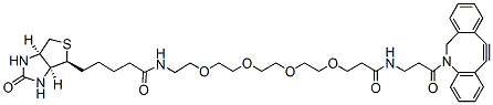
DBCO-PEG4-biotin is a click chemistry biotinylation that can enable copper-free click chemistry with azide molecules to form a stable triazole linkage. PEG4 spacer increases aqueou solubility of the molecules conjugated to the biotin compound. Reagent grade, for research purpose. Please contact us for GMP-grade inquiries.
Detail
DBCO-PEG4-biotin is a click chemistry biotinylation that can enable copper-free click chemistry with azide molecules to form a stable triazole linkage. PEG4 spacer increases aqueou solubility of the molecules conjugated to the biotin compound. Reagent grade, for research purpose. Please contact us for GMP-grade inquiries.
Other Products
Trichomonas vaginalis TaqMan PCR Detection Kits
Product Info
Document
Product Info
Overview
Detection kits for Trichomonas vaginalis
Available in TaqMan format for analysis
Norgen’s Trichomonas vaginalis End-Point PCR Kit is designed for the detection of Trichomonas vaginalis specific DNA based on the use of end-point PCR technology. This kit is designed for research use only and not for use in diagnostic procedures. The kit includes Master Mix and primers for the specific amplification of a 335 nucleotide region of the Trichomonas vaginalis genome, as well as a positive control and a negative control to confirm the integrity of the kit reagents. In addition, the kit contains loading dye and a DNA ladder to facilitate analysis of the results.
The detection of Trichomonas vaginalis specific DNA is based on end-point PCR technology, utilizing polymerase chain reaction (PCR) for the amplification of specific Trichomonas vaginalis DNA sequences. For analysis of the PCR data, the PCR reaction is loaded on an agarose DNA gel along with the provided DNA ladder for qualitative analysis.
Storage Conditions and Product Stability All kit components can be stored for 2 years after the date of production without showing any reduction in performance.
All kit components should be stored at -20°C upon arrival. Repeated thawing and freezing (> 2 x) of the Master Mix and Positive Control should be avoided, as this may affect the performance of the assay. If the reagents are to be used only intermittently, they should be frozen in aliquots.
Propargyl-PEG3-NHS ester is a Click Chemistry reagent with a propargyl group and an NHS ester group. The propargyl group can react with biomolecules containing azide group via copper catalyzed Click Chemistry reaction. The NHS ester is an amine reactive group which can be used for derivatizing peptides, antibodies, amine coated surfaces etc. Reagent grade, for research purpose. Please contact us for GMP-grade inquiries.
Document
Propargyl-PEG3-NHS ester is a Click Chemistry reagent with a propargyl group and an NHS ester group. The propargyl group can react with biomolecules containing azide group via copper catalyzed Click Chemistry reaction. The NHS ester is an amine reactive group which can be used for derivatizing peptides, antibodies, amine coated surfaces etc. Reagent grade, for research purpose. Please contact us for GMP-grade inquiries.