
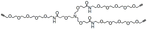
Tri(propargyl-PEG5-NHCO-ethyloxyethyl)amine is a click chemistry branched linker. The propargyl groups can react with azide-bearing molecules via copper catalyzed Click Chemistry.


Tri(propargyl-PEG5-NHCO-ethyloxyethyl)amine is a click chemistry branched linker. The propargyl groups can react with azide-bearing molecules via copper catalyzed Click Chemistry.

Gly-Gly-Gly-PEG4-DBCO is a cleavable PEG linker used in the synthesis of antibody-drug conjugates. Reagent grade, for research purpose. Please contact us for GMP-grade inquiries.

Gly-Gly-Gly-PEG4-DBCO is a cleavable PEG linker used in the synthesis of antibody-drug conjugates. Reagent grade, for research purpose. Please contact us for GMP-grade inquiries.

Propargyl-PEG7-t-butyl ester comprises a propargyl group and a t-butyl protected carboxyl group. The propargyl group reacts with azide compounds via copper catalyzed Click Chemistry to form a stable triazole linkage. Under acidic conditions, the carboxyl group can be deprotected. The hydrophilic PEG units enhance solubility of the molecule in aqueous media. Reagent grade, for research purpose. Please contact us for GMP-grade inquiries.

Propargyl-PEG7-t-butyl ester comprises a propargyl group and a t-butyl protected carboxyl group. The propargyl group reacts with azide compounds via copper catalyzed Click Chemistry to form a stable triazole linkage. Under acidic conditions, the carboxyl group can be deprotected. The hydrophilic PEG units enhance solubility of the molecule in aqueous media. Reagent grade, for research purpose. Please contact us for GMP-grade inquiries.

Description
The PM2510 ExcelBand™ Enhanced 3-color Regular Range Protein Marker is a ready-to-use three-color protein standard with 10 pre-stained proteins covering a wide range of molecular weights from 10 to 180 kDa in Tris-Glycine Buffer (9 to 170 kDa in Bis-Tris (MOPS) buffer and 10 to 170 kDa Bis-Tris (MES) buffer). Proteins are covalently coupled with a blue chromophore except for two reference bands (one green and one red band at 25 kDa and 75 kDa respectively) when separated on SDS-PAGE (Tris-Glycine buffer). PM2510 ExcelBand™ Enhanced 3-color Regular Range Protein Marker is designed for monitoring protein separation during SDS-polyacrylamide gel electrophoresis, verification of Western transfer efficiency on membranes (PVDF, nylon, or nitrocellulose) and for approximating the size of proteins.
Features
Contents
Approximately 0.2~0.6 mg/ml of each protein in the buffer (20 mM Tris-phosphate (pH 7.5), 2% SDS, 3.6 M urea, and 15% (v/v) glycerol).
Quality Control
Under suggested conditions, PM2510 ExcelBand™ Enhanced 3-color Regular Range Protein Marker resolves 10 major bands in 15% SDS-PAGE (Tris-Glycine buffer, MOPS, and MES buffer) and after Western blotting to nitrocellulose membrane.
Storage
4°C for 3 months
-20°C for long term storage
The PM2510 ExcelBand™ Enhanced 3-color Regular Range Protein Marker is a ready-to-use three-color protein standard with 10 pre-stained proteins covering a wide range of molecular weights from 10 to 180 kDa in Tris-Glycine Buffer (9 to 170 kDa in Bis-Tris (MOPS) buffer and 10 to 170 kDa Bis-Tris (MES) buffer). Proteins are covalently coupled with a blue chromophore except for two reference bands (one green and one red band at 25 kDa and 75 kDa respectively) when separated on SDS-PAGE (Tris-Glycine buffer). PM2510 ExcelBand™ Enhanced 3-color Regular Range Protein Marker is designed for monitoring protein separation during SDS-polyacrylamide gel electrophoresis, verification of Western transfer efficiency on membranes (PVDF, nylon, or nitrocellulose) and for approximating the size of proteins.