Convenient optimized on-column DNase treatment using Norgen’s RNA Purification Kits
Also includes protocol for digestion in-solution followed by RNA Clean-Up
Guaranteed RNase-Free
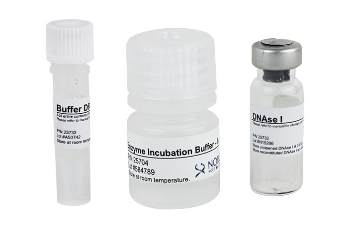
Includes Enzyme Incubation Buffer
Cat. 25710 contains one vial of 1,600 units and Cat. 25720 contains 4 vials (1,600 units/vial)
Norgen’s RNA purification kits isolate total RNA with minimal amounts of genomic DNA contamination. However, for some sensitive downstream applications, it may be desirable to remove all traces of residual DNA. Norgen’s RNase-free DNAse I Kit, with Enzyme Incubation Buffer, can be used for optional on-column DNase digestion with any of Norgen’s RNA purification kits. Alternatively, after isolating total RNA using one of Norgen’s RNA purification kits, the RNA elution can be treated with this DNase I. The RNA can then be purified from the DNase using Norgen’s RNA Clean-Up and Concentration Kit (Cat# 23600), and the RNA can then be used in downstream applications.
Details
Each RNase-Free DNase I Kit is supplied complete with sufficient enzyme and enzyme incubation buffer for 50 or 200 reactions.
Storage Conditions The DNase I provided is in lyophilized form. It is stable for at least 3 months if stored at room temperature. However, it is recommended to store the DNase I vial at 2 – 8ºC (or below) upon receipt to maintain stability beyond 3 months. Buffer DR and Enzyme Incubation Buffer can be stored at room temperature. After reconstitution with Buffer DR (see product manual), the DNase I should be stored at -20ºC. All reagents should remain stable for at least 1 year in their unopened containers at the appropriate storage temperature.
Component
Cat. 25710 (50 rxns)
Cat. 25720 (200 rxns)
DNase I
1 vial / 1,600 units
4 vials (1,600 units/vial)
Buffer DR
1 mL
4 x 1 mL
Enzyme Incubation Buffer
6 mL
4 x 6 mL
Product Insert
1
1
Other Products
Propargyl-PEG4-NHS ester
Product Info
Document
Product Info
Propargyl-PEG4-NHS ester is an amine reactive PEG linker with an alkyne group. Under the copper catalyzation, alkyne group can react with azide-bearing biomolecules to form a stable triazole linkage. The hydrophilic PEG spacer increases solubility in aqueous media. Reagent grade, for research purpose. Please contact us for GMP-grade inquiries.
Document
Propargyl-PEG4-NHS ester is an amine reactive PEG linker with an alkyne group. Under the copper catalyzation, alkyne group can react with azide-bearing biomolecules to form a stable triazole linkage. The hydrophilic PEG spacer increases solubility in aqueous media. Reagent grade, for research purpose. Please contact us for GMP-grade inquiries.
Biocolor’s Purple-Jelley assay kit is the perfect tool for accurate measurement of hyaluronic acid / Hyaluronan levels in your samples. This colorimetric assay is optimised for quantitative analysis in-vivo, tissue-derived hyaluronic acid / Hyaluronan and includes full step-by-step instructions.
Colorimetric Detection (655nm) (Endpoint)
Hyaluronic Acid: A gentle giant!
Hyaluronic acid, in its hydrated form, is a unique carbohydrate polymer, often referred to as a ‘gentle giant.’ It consists of a lengthy, flexible, non-branching chain with a repeating disaccharide pattern. This disaccharide is composed of alternating uronic acid and aminosugar units.
Why is our kit called ‘Purple-Jelley’?
Discovering the J-Aggregate Effect in Cyanine DyesIn 1936, Edwin Jelley made a fascinating observation, documented it in a letter to Nature (Nature 138, 1009 – 1010). He noted a peculiar behaviour of certain cyanine dyes, that when dissolved in 5 M NaCl, they dyes exhibited a third absorbance peak at a longer wavelength, around 650nm. In deionized water, however, they displayed only a double peak at approximately 540nm and 570nm. The 650nm peak in concentrated dye solutions resulted from the aggregation of dye molecules and was later termed a ‘J-aggregate,’ in honor of Edwin Jelley. The J-aggregate is known as a supra-molecular complex, formed by stacking individual dye molecules.
Subsequent research in the 1960s, notably by Kay et al. (J. Physical Chem. 68, 1896 – 1906), revealed that various biological polymers, including proteins, DNA, polar lipids, and glycosaminoglycans, could also induce this third absorbance peak. This phenomenon led to the development of the Purple-Jelley assay, named after the purple color of the dye reagent and Edwin Jelley himself.
An overview of the Purple-Jelley assay steps:
During the assay, hyaluronic acid is selectively purified during the assay sample preparation protocol. This is then reacted with the Purple-Jelley dye reagent, and the absorption of the characteristic third wavelength recorded. By comparison with a calibration curve the hyaluronic acid content of the sample can be measured.
Step 1. The assay protocol takes tissue samples through a sequential sample preparation protocol which involves enzymatic protein digestion, followed by precipitation and purification of GAGs, culminating in the precipitation of purified Hyaluronic acid.
Step2. The processed sample is then incubated for 10 minutes with the Purple-Jelley dye reagent, forming a coloured product which can be measured spectrophotometrically.
Step 3. The Hyaluronic acid content of unknown samples can be calculated by comparison against a calibration curve prepared using a standard comprising hyaluronic acid (supplied with the kit).
Assay range
10 – 100µg/ml
Limit of Detection
10µg/ml
Detection Method
Colorimetric Detection (655nm) (Endpoint)
Measurements per kit
100 in total (allows a maximum of 46 samples to be run in duplicate alongside a standard curve).
Suitable Samples
In-vivo: Hyaluronic acid purified from in-vivo tissues. The kit protocol involves extraction and purification of hyaluronic acid prior to reaction with the Purple-Dye reagent.
Precautions
This kit is designed for research use only. Not for use in diagnostic procedures. Kit requires access to a centrifuge, as well as a spectrophotometer/colorimeter capable of colorimetric, absorbance detection at 655nm. Specific sample preparation protocols may require customer to provide further reagents, consult assay manual for further information.
2. Hyaluronan Reference Standard (1x 5ml, 0.2mg/ml soluble Hyaluronic Acid)
3. Precipitating Reagent (2x 34ml)
4. Sodium Chloride (1x 20ml)
5. Cetylpyridinium Chloride (1x 20ml)
6. TRIS-buffered Saline (5x tablets)
7. 2ml screw-cap tubes for preparation of samples.
8. Assay kit manual
NB: Additional reagents may be required for sample preparation prior to assay. Consult manual or contact us for further details.
Document
Biocolor’s Purple-Jelley assay kit is the perfect tool for accurate measurement of hyaluronic acid / Hyaluronan levels in your samples. This colorimetric assay is optimised for quantitative analysis in-vivo, tissue-derived hyaluronic acid / Hyaluronan and includes full step-by-step instructions.
AAV Purification from any input – cell fraction or media fraction
High AAV recovery, up to 90%
No specialized equipment needed
Purification from a variety of AAV serotypes (including AAV6 and AAV9)
Yields highly active AAV for in vivo and in vitro experiments
Purification is based on spin column chromatography that uses Norgen’s resin separation matrix
Recombinant adeno-associated virus (AAV) vectors are highly promising tools for both in vitro and in vivo gene transfer. Norgen’s AAV Purification Kits provide fast and simple procedures for concentrating and purifying AAV vectors from cell lysate and cell culture media. Purification is based on precipitation onto Norgen Biotek’s proprietary resin. Contaminating cellular debris is largely removed from the sample via a centrifugation step, while contaminating DNA and RNA is reduced using enzymatic digestion. AAV vector purified in this manner is highly active for use in in vitro and in vivo transduction experiments.
AAV Purification Kit
Norgen’s AAV Purification Kit contains sufficient materials for 15 preparations (33.5 mL per prep of supernatant (SN) or a total of 500 mL of supernatant input). Approximately 1 mL of cell pellet can be purified per prep, up to a maximum of 15 mL of cell pellet in total for the entire kit. Up to 33X sample concentration.
AAV Purification Mini Kit
Each spin column is able to concentrate and purify AAV from 0.5-8 mL of cell pellet, cell culture media, or cells and culture media mixed together. Up to 50X sample concentration. AAV vector purified in this manner is highly active for use in in vitro transduction experiments, and is eluted into a small volume (200 µL). Preparation time for 4 samples is 1.5 hours, with 45 minutes of hands-on time.
AAV Purification Midi Kit
Each spin column is able to concentrate and purify AAV from 8 mL up to 45 mL of input consisting of cell pellet, cell culture media, or cells and culture media mixed together. Up to 50X sample concentration. AAV vector purified in this manner is highly active for use in in vitro transduction experiments, and is eluted into a small volume (1 mL). The kit may be used to purify up to 8 x 25 mL or 4 x 45 mL of samples using the included columns. Preparation time for 4 samples is approximately 2 to 2.5 hours, with 1.5 hours of hands on time.
AAV Purification Maxi Kit (Slurry Format)
Each spin column is able to concentrate and purify AAV from 45 mL to 90 mL of input consisting of cell pellet, cell culture media, or cells and culture media mixed together. Up to 200X sample concentration. AAV vector purified in this manner is highly active for use in in vitro transduction experiments, and is eluted into a small volume (1-10 mL) using the optional concentration step. The kit may be used to purify up to 1 x 900 mL samples or 10 x 45-90 mL samples using the included columns. Preparation time for 1 x 900 mL sample is approximately 2.5 to 3.5 hours, with an optional concentration step requiring an additional 30 min.
At least 1 x 10¹⁰ AAV particles as determined by qPCR
AAV Vector Serotype
Any
Average Recovery
> 80%
Input Type
Cells, media, or mixed
Input Volume
8 mL – 45 mL
Minimum Elution Volume
1 mL
Time to Complete 4 Purifications
2 – 2.5 hours
Storage Conditions and Product Stability DNAse I and RNAse A should be stored at -20°C upon arrival. Elution Buffer O should be stored tightly capped at 4°C upon arrival. All other solutions should be kept tightly sealed and stored at room temperature. Once opened, the solutions should be stored at 4°C. This kit is stable for 1 year after the date of shipment.
Component
Cat. 66100 (15 preps)
Cat. 63200 (20 preps)
Cat. 63300 (4-8 preps)
Cat. 63250 (1-10 preps)
Lysis Buffer S
5.5 mL
5.5 mL
5.5 mL
20 mL
DNAse I
–
2 x 25 uL
2 x 25 uL
210 μL
RNAse A
–
60 μL
60 μL
240 μL
HL-SAN Nuclease
102 μL
–
–
–
Binding Buffer A
20 mL
4 mL
4 mL
2 x 8 mL
Purification Solution C
60 mL
–
–
–
Purification Solution D
130 mL
–
–
–
Wash Solution C
2 x 130 mL
60 mL
60 mL
3 x 60 mL
Slurry E
12.5 mL
–
–
2 x 14.5 mL
Elution Buffer O
66 mL
8.5 mL
8.5 mL
66 mL
Protein Neutralizer
4 mL
4 mL
4 mL
4 mL
Spin Columns
–
20
–
–
Mini Spin Columns
–
20
–
–
Midi Spin Columns (grey contents) with Collection Tubes
–
–
8
10
Midi Spin Columns (white contents) with Collection Tubes
–
–
8
–
Maxi Spin Columns (grey contents) with Collection Tubes
–
–
–
10
Maxi Spin Columns (white contents) with Collection Tubes