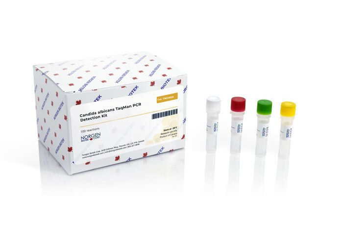
CE-IVD marked version available for in vitro diagnostic use
Available in TaqMan format for analysis
Candida albicans is an opportunistic yeast and is the most common fungal pathogen found in the human body. C. albicans can be detected in the gastrointestinal tract, mouth, and vagina of approximately half of healthy adults. It is typically a commensal organism and makes up part of the natural human microflora; however it can overgrow and become pathogenic as a result of various conditions. For example, individuals who have taken recent courses of antibiotics, have a weakened immune system or have diabetes have an increased risk of developing a Candida albicans infection. When an overgrowth occurs, this can lead to common infections such as urinary tract infections, genital yeast infections, oral thrush, and mucocutaneous candidiasis. In the more severe cases, when Candida albicans enters the bloodstream or organs, it can lead to loss of sight, blood and bone infections, endocarditis, meningitis or inflammation of the intraabdominal lining.
Storage Conditions and Product Stability All kit components can be stored for 2 years after the date of production without showing any reduction in performance.
All kit components should be stored at -20°C upon arrival.
Apoptosis is an essentially normal physiological process that removes now redundant, cells, particularly during embryonic development and early growth. In adult animals the process removes cells that are irreparable. The apoptotic process is also involved in many major diseases such as cancer, where transformed tumour cells have their apoptotic process disabled, permitting cell cycling to continue unchecked. In contrast some forms of senile dementia may result from excessive apoptotic induction of neural cells.
The apoptotic process in mammalian cells is a rapid event (2‐4 hours). Within this short time span an apparently viable cell can be quietly dismantled, to disappear leaving no visible trace of its former existence.
How is apoptosis detected or measured?
An apoptosis cascade of activators, effectors and regulators has been identified. This in turn led to a range of apoptosis assays being devised to detect and monitor these events. Some laboratories will employ two distinct assays, one selected to detect early (initiation) apoptotic events, while a second assay will target a later (execution) event. Apoptosis assays, based on methodology, can be classified into four major inter‐linked groups:
[1] DNA fragmentation (electrophoresis and nick end labelling, TUNEL).
[2] Apoptotic proteases (fluorescently labelled antibodies to the caspases).
[3] Flow cytometric analysis (FACS, incorporating other group assays).
Biocolor’s APOPercentage assay is based on the latter. Further information can be found under the ‘Mode of Action’ Tab.
How does APOPercentage detect apoptosis?
The mammalian cell membrane has been described as a semi‐fluid mosaic structure, composed of phospholipids with a diverse group of inserted proteins and some cholesterol. The phospholipids are the major components of the membrane and are arranged in the form of a ‘bi‐layer’; which is asymmetric in composition, structure, and function.
To ensure normal transmembrane functions the phospholipids must be maintained in an asymmetric composition. The process is regulated by ‘flippases’, which catalyse the active transport of aminophospholipids from the outer to inner monolayer. However, in cells undergoing apoptosis, flippase is overwhelmed by the action of another enzyme, termed ‘floppase’ or ‘scramblase’. The net effect is a scrambling of the phospholipid distribution between the inner and outer monolayers.
Cell membrane changes during apoptosis
The APOPercentage assay utilises an intense, pink-coloured dye reagent which is taken up during in-vitro culture by apoptosis-committed cells. This uptake occurs at the stage of Phosphatidylserine transmembrane movement, as produced by the flipflop mechanism. Dye uptake continues until blebbing occurs. No further dye can then enter the now defunct cell and the dye that has accumulated within the cell is not released (unlike necrotic cells which release dye).
Since the dye reagent is excluded or not retained by healthy or necrotic cells it therefore acts as a specific label for apoptotic cells.
How are APOPercentage-labelled cells quantified?
Labelled apoptosis cells may then by conveniently analysed by the following methods:
Direct Analysis The intense pink colour of the labelled cells can be visually assessed using brightfield microscopy. Apoptosis in substrate-adherent cell populations is therefore readily quantified using image analysis techniques. This technique is the most sensitive with the ability of detecting one single apoptotic cell per well.
Colorimetry protocol Dye that accumulates within apoptotic cells is released into solution via addition of Dye Release Reagent. The concentration of this intracellular dye is then measured at 550nm using a microplate colorimeter/spectrophotometer.
NB: The APOPercentage assay kit does NOT require the use of a Flow Cytometer.
Limit of Detection
A single cell (via image analysis method)
Detection Method
Colorimetric (550nm) (Endpoint) or Image Analysis based
Measurements per kit
Sufficient for 4×24 well plates or 6×96 well plates
Suitable Samples
Adherent mammalian cells (in-vitro)
APOPercentage kit contents:
1. APOPercentage Dye (1x5ml)
2. Dye Release Reagent (1x150ml)
3. Phosphate Buffered Saline (PBS) (1x120ml)
4. 24-well starter plate.
5. Assay kit manual.
The Colorimetric Protocol requires a Microplate Colorimeter / Spectrophotometer.
Additional 96-well plates will be required for use when reading dye absorbance values.
The Direct Detection Protocol Requires an inverted stage microscope with an attached digital camera.
NB: Additional reagents (typically culture medium and suitable apoptosis treatments) may be required for sample preparation prior to assay. Consult manual or contact us for further details.
Document
The APOPercentage™ Apoptosis kit is a dye-based, colorimetric assay for detection and measurement of apoptosis (programmed cell death) during in-vitro cell culture.
NGS Cell Free DNA Library Prep Kit (illumina and MGI Platforms)
Product Info
Document
Product Info
The NGS Cell Free DNA Library Prep Kit (illumina and MGI Platforms) was developed for the construction of high quality cell-free DNA (cfDNA) libraries using 1 ng to 50 ng of cell-free DNA as input. The kit has a simple work flow and a fast procedure. Multiplexing of the cell free DNA library is possible based on the index type.
NGS Cell Free DNA Library Prep Kit Workflow
The main source of cell-free DNA (cfDNA) is derived from apoptotic hematopoietic cells in blood and found in the plasma. The length of the cfDNA is about 150-200 bp in length. Circulating tumor DNA (ctDNA) derived from malignant tumors is a part of cfDNA. Both cfDNA and ctDNA can be used as a noninvasive biomarker since it offers a better approach in comparison to invasive tissue biopsies.
NGS has been used for cfDNA and ctDNA sequencing in the field of liquid biopsy as it provides a whole genome level of molecular profiling. One of the hurdles for cfDNA sequencing is the difficulty of library preparation from the limited amount of cfDNA obtained from plasma. Our cell-free NGS kit makes it easy to get enough libraries from limited input in just 1.5 hours.
Three index types are available for the NGS Cell Free DNA Library Prep Kit of the illumina platform:
Non-index (Cat.# 30029): Libraries do not have index.
Index(Cat.# 30031): Each of our index primers contains a unique 6-base index sequence that can be used for sample identification. Total 48 library multiplexing is possible. Index information can be downloaded here.
Unique dual index (Cat.# 30033): Cell-free DNA library multiplexing up to 96 samples is possible with the unique dual indexes. We have developed a Four-Base Difference Index System. The system have at least 4 bases different from each other in the 8 bases index length. The primers effectively minimize sequencing errors such as mis-assignment, index hopping, index contamination etc. Index information can be downloaded here.
Indexes are available for the MGI platform kits (Cat.# 34031).
Kit advantages:
Fast and simple protocol
Hands-on time is around 10 minutes
The total protocol time is only 1.5 hours
Easy procedure based on:
Ready-to-use master mix for easy setup of reactions
Less reaction components to simplify bench work
Less magnetic beads used for cleanup procedures: This can reduce more than 50% of the beads consumption
Guaranteed high library conversion efficiency
Low input cell-free DNA: From 1 ng to 50 ng
Comparison of library conversion efficiency with different samples under the same condition.
Comparison of library yield with different samples under the same condition.
Document
The NGS Cell Free DNA Library Prep Kit (illumina and MGI Platforms) was developed for the construction of high quality cell-free DNA (cfDNA) libraries using 1 ng to 50 ng of cell-free DNA as input. The kit has a simple work flow and a fast procedure. Multiplexing of the cell free DNA library is possible based on the index type.