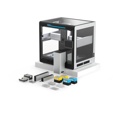
Opentrons Flex Magnetic Bead Protein Purification Workstation
Facebook
X
Pinterest
Email
For scaling up and fully automating magnetic bead-based protein purification workflows.
With the Flex Protein Purification Workstation, you can automate your small-scale protein purification and proteomics sample processing at the scale you need to increase efficiency, reduce errors, and save hands-on time. This Opentrons Protein Purification Workstation uses magnetic beads for purification of proteins.
Optional add-ons can be purchased at a 10% discount when ordered with the Flex Magnetic Bead Protein Purification Workstation*
Detail
For scaling up and fully automating magnetic bead-based protein purification workflows.
With the Flex Protein Purification Workstation, you can automate your small-scale protein purification and proteomics sample processing at the scale you need to increase efficiency, reduce errors, and save hands-on time. This Opentrons Protein Purification Workstation uses magnetic beads for purification of proteins.
Optional add-ons can be purchased at a 10% discount when ordered with the Flex Magnetic Bead Protein Purification Workstation*
Other Products
R6628 MagPure Serum miRNA Kit
Product Info
Document
Product Info
Introduction
The kit offers the unique feature to isolate total RNA including small RNA and DNA from serum and plasma without the need to resort to the cumbersome phenol/chloroform extraction or a time consuming proteinase digest. RNA purified using the kit is ready for applications such as RT-PCR, Northern blotting, poly A+ RNA (mRNA) purification, nuclease protection, and in vitro translation.
Details
Specifications
Features
Specifications
Main Functions
Isolation miRNA from 0.3-0.5ml serum or plasma using magnetic particles
Applications
RT-PCR, cDNA synthesis, second generation sequencing
Purification method
Polydisperse magnetic beads
Purification technology
Magnetic beads technology
Process method
Manual or automatic
Adaptive instrument
Nucleic acid extractor, pipetting workstation
Sample type
Serum, plasma, acellular samples
Sample amount
300μl
Principle
This product is based on the purification method of high binding magnetic particles. The sample material is denatured in Lysis Buffer. The protein is then precipitated by Protein Precipitation Solution and pelleted by centrifugation. After adding magnetic particles and binding solution, RNA will be adsorbed on the surface of magnetic particles, and impurities such as proteins will be removed without adsorption.The adsorbed particles were washed with washing solution to remove proteins and impurities, washed with ethanol to remove salts, and finally RNA was eluted by Elution Buffer.
Advantages
Safe – no phenol chloroform extraction and no use of Trizol reagent
Fast – several samples can be extracted in 60 minutes by column method
Kit Contents
Contents
R662801
R662802
R662803
Purification Times
48 Preps
96 Preps
5 x 96 Preps
MagPure Particles N
1.7 ml
3.5 ml
17 ml
Buffer CFL
6 ml
12 ml
60 ml
Buffer CPL
1.8 ml
3.5 ml
20 ml
Buffer MGW1*
30 ml
60 ml
250 ml
Buffer MW2*
12 ml
25 ml
100 ml
RNase Free Water
10 ml
20 ml
60 ml
Storage and Stability
MagPure Particle N should be stored at 2–8°C upon arrival. However, short-term storage (up to 8 weeks) at room temperature (15–25°C) does not affect their performance. The remaining kit components can be stored at room temperature (15–25°C) and are stable for at least 18 months under these conditions.
Document
The kit offers the unique feature to isolate total RNA including small RNA and DNA from serum and plasma without the need to resort to the cumbersome phenol/chloroform extraction or a time consuming proteinase digest. RNA purified using the kit is ready for applications such as RT-PCR, Northern blotting, poly A+ RNA (mRNA) purification, nuclease protection, and in vitro translation.
Trichinellosis is caused by Trichinella spiralis, a parasitic nematode of pork, horse and wild animals. Humans can be infected by eating raw or undercooked meat from infected animals. Most infected people do not show any symptoms. However, in some cases, symptoms appear during intestinal stage (diarrhea and abdominal pain) and muscular stage (muscle pain, fever, edema). Diagnosis is based on signs and symptoms, but in many cases it is not conclusive for the diagnosis. Serological assays have an important place on the final diagnosis of the disease. The ELISA kit has shown appropriate performance for the serological screening of trichinellosis.
This product is manufactured by Bordier Affinity Products in Switzerland and distributed in Germany exclusively by Milenia Biotec.
Method/Platform
ELISA in microplate format
Range/Assay Sensivity
95% sensitivity, 98% specificity
Test Principle
The kit provides all the material needed to perform 96 enzyme-linked immunosorbent assays (ELISA) on breakable microtitration wells sensitized with Trichinella spiralisexcreted/secreted (E/S) larval antigens. Specific antibodies in the sample will bind to these antigens and washing will remove unspecific antibodies. The presence of parasite specific antibodies is detected with a Protein A – alkaline phosphatase conjugate. A second washing step will remove unbound conjugate. Revealing bound antibodies is made by the addition of pNPP substrate which turns yellow in the presence of alkaline phosphatase. Color intensity is proportional to the amount of Trichinella spiralis specific antibodies in the sample. Potassium phosphate is added to stop the reaction. Absorbance at 405 nm is read using an ELISA microplate reader. The test can be performed with automatic systems, but this must be validated by the user.
Trichinellosis is caused by Trichinella spiralis, a parasitic nematode of pork, horse and wild animals. Humans can be infected by eating raw or undercooked meat from infected animals. Most infected people do not show any symptoms. However, in some cases, symptoms appear during intestinal stage (diarrhea and abdominal pain) and muscular stage (muscle pain, fever, edema). Diagnosis is based on signs and symptoms, but in many cases it is not conclusive for the diagnosis. Serological assays have an important place on the final diagnosis of the disease. The ELISA kit has shown appropriate performance for the serological screening of trichinellosis.
The ExcelTaq™ 5X Fluorescent PCR Master Mix is a ready-to-use mixture for amplifying targeted DNA fragments. It is designed to serve as a master mix for virtually all PCR applications. This product is supplied as a 5X concentrated mixture containing all the essential ingredients for PCR with the exception of templates and primers. In addition, the mixture contains electrophoresis tracking dye (Bromophenol blue) and a safer fluorescent DNA staining dye, which enables the user to track the electrophoresis process in real time as well as eliminates the need for the staining process. The resultant PCR reaction mixture is sufficiently dense enough to be loaded directly into a TAE or TBE buffered gel for electrophoresis.
Features
5’→3’ DNA polymerase activity
No detectable 3’→5′ exonuclease (proofreading) activity
Generates PCR products with 3′-dA overhangs
High yield PCR
High reproducibility
Reduced pipetting errors
For direct loading into electrophoresis analysis
DNA bands can be visualized directly under UV or 470 nm blue light illumination
Applications
Routine PCR
Colony PCR
High throughput PCR
Amplification of DNA fragments up to 8 kb
Generation of PCR products for TA cloning
DNA labeling
Storage
Protected from light and avoid multiple freeze/thaw cycles 4°C for 6 months -20°C for 24 months
Document
The ExcelTaq™ 5X Fluorescent PCR Master Mix is a ready-to-use mixture for amplifying targeted DNA fragments. It is designed to serve as a master mix for virtually all PCR applications. This product is supplied as a 5X concentrated mixture containing all the essential ingredients for PCR with the exception of templates and primers. In addition, the mixture contains electrophoresis tracking dye (Bromophenol blue) and a safer fluorescent DNA staining dye, which enables the user to track the electrophoresis process in real time as well as eliminates the need for the staining process. The resultant PCR reaction mixture is sufficiently dense enough to be loaded directly into a TAE or TBE buffered gel for electrophoresis.