
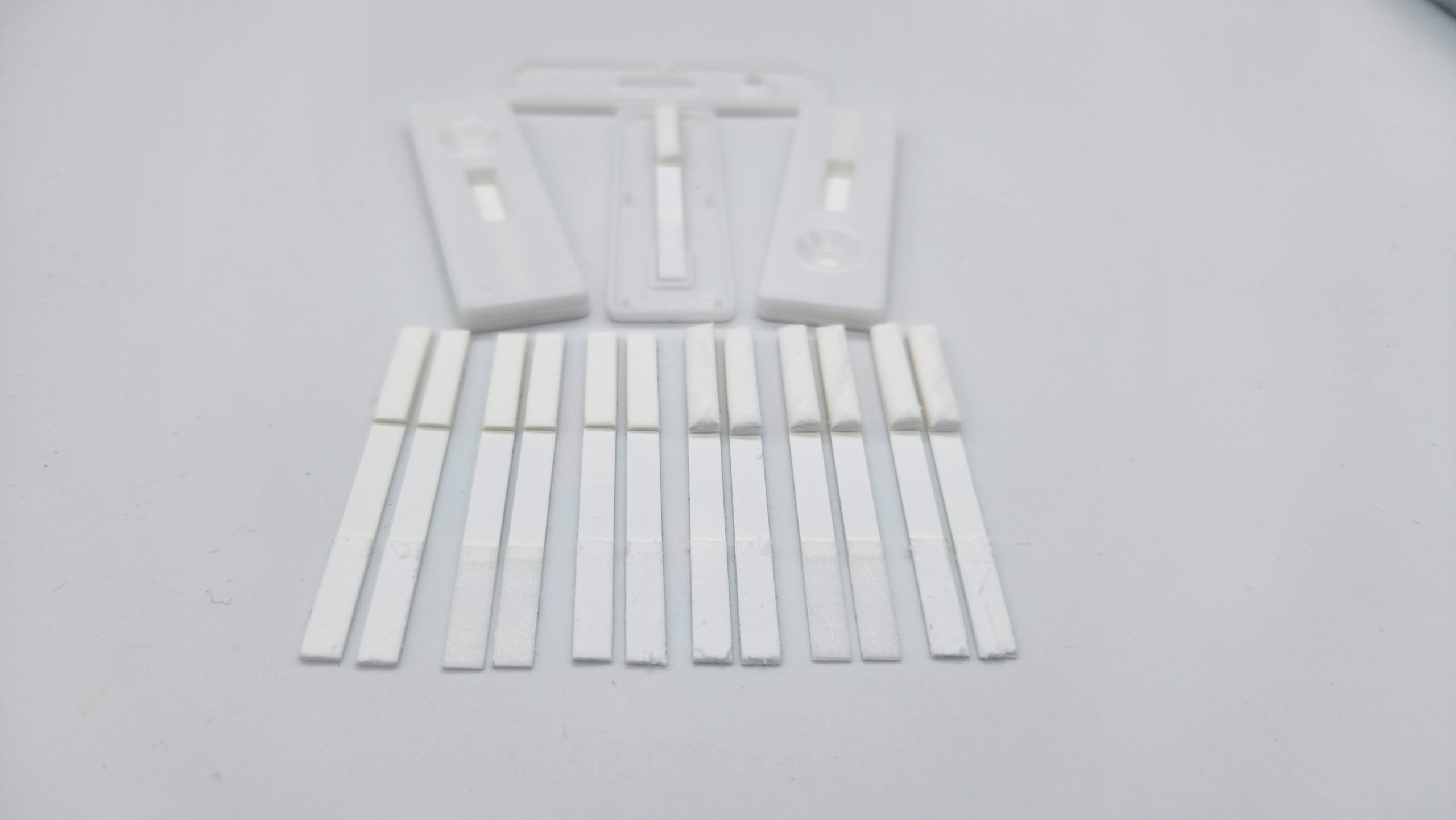

- Twenty 4.5X60mm strips of each combination of nitrocellulose (4X), wicking material (2X), and sample pad (3X)
- A total of 480 strips are provided
- 100 cassettes
- 125mL of buffer
Description
Blank lateral flow strips are the perfect companion during the development and feasibility testing of a lateral flow device. IN THE BEGINNING, the capture and test line reagents can be spotted onto the nitrocellulose portion of the blank strips at different concentrations, volumes and buffer conditions. This enables the user to determine the optimal conditions for subsequent spraying and enables the user to verify the test and controls will respond according to expectations before scaling up the process.
These blank strips are compatible with all of our colloidal gold products AND:
Lateral flow cassettes: C02022
Streptavidin CdSe/ZnS Quantum Dots For Lateral Flow: AU2043
Streptavidin Europium Chelate Microspheres For Lateral Flow: AU2050
Streptavidin Colloidal Gold For Lateral Flow: AU2017
RNase-Free Lateral Flow Assay Running Buffer Screening Pack: AU2035


