DNase activity in a convenient and sensitive lateral flow colormetric assay that delivers results in real time. Great for Quality Testing for DNase contamination of materials and supplies
Detail
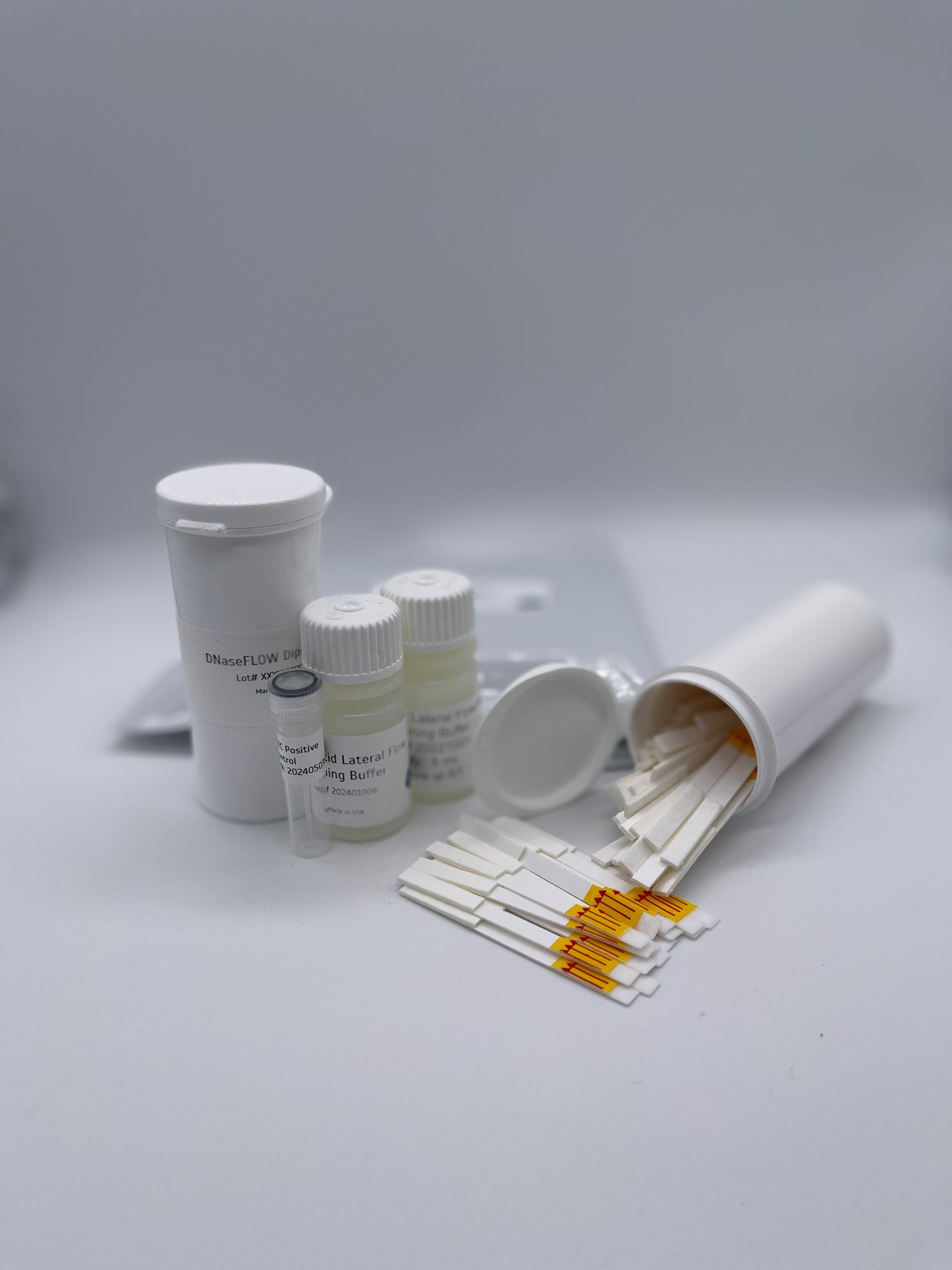
DNase activity in a convenient and sensitive lateral flow colormetric assay that delivers results in real time. Great for Quality Testing for DNase contamination of materials and supplies.
Attogene’s DNaseAlarm Lateral Flow test is designed for the sensitive and accurate analysis of DNAse activity in liquid samples. DNase Alarm uses a synthetic DNA substrate that attaches to the streptavidin colloidal reporter molecule (gold) using a 5’ biotin. The DNA substrate also contains a FAM molecule that enables it to be captured by the anti-FAM antibody (test line). In the absence of DNases, the DNA oligo tethers gold to the test line giving a visual test line. When DNases are present, the DNA substrate is degraded, and the gold particles can no longer be tethered to the test line thus, signal is lost. Since the cleavage of the DNA Substrate increases over time when DNase activity is present, results can be evaluated kinetically. This assay has applications for quality control testing and analysis of unit activities of DNase and DNase inhibitors. DNase’s can cause havoc in laboratories working with DNA and are important to perform routine testing.
This test can be used to rapidly and efficiently detect DNase’s in both liquid and on solid surfaces and a perfect tool for monitoring manufacturing.
Other Products
EXTRAClean Urine Exosome Purification and RNA Isolation Kits
Product Info
Document
Product Info
Overview
Ensure optimal results for sensitive applications like NGS
Up to a tenfold increase in microRNA mapping during sequencing runs to reduce costs
Purification and enrichment of intact urinary exosomes for functional studies.
Bind and elute all RNA irrespective of size or GC content, without bias.
Isolate all sizes of RNA, including microRNA, irrespective of size or GC content, without bias.
No phenol extractions, Proteinase K treatment, nor carrier RNA required.
No time-consuming ultracentrifugation, filtration nor special syringes are required.
No precipitation reagents nor overnight incubation required.
Compatible with urine from any species.
Pure exosomes are purified and are free-from any other RNA-binding proteins.
Purification is based on EXTRAClean spin column chromatography that uses Norgen’s proprietary resin separation matrix.
Norgen’s EXTRAClean Urine Exosome Purification and RNA Isolation Mini Kit constitutes an all-in-one system for the purification of exosomes and the subsequent isolation of RNA from different urine sample volumes ranging from 250 μL to 1 mL. The purification is based on spin column chromatography that employs Norgen’s proprietary resin. The EXTRAClean columns undergo stringent processing and rigorous quality control measures to minimize contamination traces, ensuring optimal results for sensitive applications such as NGS. The kit is designed to isolate all sizes of RNA, including microRNA. The kit provides a clear advantage over other available kits in that it does not require any special instrumentation, protein precipitation reagents, extension tubes, phenol/chloroform or protease treatments. Moreover, the kit allows the user to elute into a flexible elution volume ranging from 50 μL to 100 μL. The purified RNA is free from any protein-bound circulating RNA and of the highest integrity. The purified RNA can be used in a number of downstream applications including real time PCR, reverse transcription PCR, NGS application, Northern blotting, RNase protection and primer extension, and expression array assays.
All sizes, including miRNA and small RNA (< 200 nt)
Elution Volume
50-100 μL
Time to Complete 10 Purifications
35-40 minutes
Average Yields
Variable depending on specimen
Storage Conditions and Product Stability All buffers should be kept tightly sealed and stored at room temperature. This kit is stable for 2 years after the date of shipment. It is recommended to warm Lysis Buffer A for 20 minutes at 60°C if any salt precipitation is observed.
Bis-propargyl-PEG6 has two propargyl groups which can participate in Click Chemistry reactions to yield a stable triazole linkage with azide compounds; copper is needed as a catalyst. The 6 units of PEG increase the hydrophilicity of the compound. Reagent grade, for research purpose. Please contact us for GMP-grade inquiries.
Document
Bis-propargyl-PEG6 has two propargyl groups which can participate in Click Chemistry reactions to yield a stable triazole linkage with azide compounds; copper is needed as a catalyst. The 6 units of PEG increase the hydrophilicity of the compound. Reagent grade, for research purpose. Please contact us for GMP-grade inquiries.
Magen’s HiPure columns are prepared by high quality glass fiber filter membrane as raw materials through membrane cutting, membrane release, ring release, ring pressing, gland, weighing and other processes. HiPure nucleic acid adsorption columns have the characteristics of long-term stability and high binding capacity. Experiments show that the highest binding capacity and binding efficiency of HiPure nucleic acid adsorption columns are basically unchanged when stored at room temperature for 4 years.
The series of nucleic acid columns produced by Magen Biotech are based on carefully selected imported glass fiber membranes (GF/B, GF/D, GF/F). Columns production processes such as polypropylene injection molding materials, injection molding process, and downstream membrane packing and compression rings are strictly controlled. This is to ensure that the column has extremely high adsorption capacity and long-term stability. Compared with conventional products on the market, Magen’s columns are with varieties, and binding rate will not change when stored at room temperature for 4 years.
Details
Specifications
Features
Specifications
Recommended application
gDNA extraction, RNA extraction, DNA/RNA clean up
Preservation conditions
Room temperature
Stability
Up to 4 years
Filter membrane
High quality glass fiber filter GF/B, 2 layers
Membrane aperture
1.0μm
Maximum binding yield of plasmid
15 μg
Maximum yield of alcohol mediated Binding
100 μg
Single liquid carrying capacity of column
700 μl
Minimum elution volume
30 μl
Withstand centrifugal force
12,000 x g
Centrifuge
Small high speed centrifuge (2ml)
Adsorption Mechanism
Based on the negatively charged DNA skeleton, it has a high affinity for positively charged glass fibers. In high salt and ethanol solutions, DNA/RNA binds to glass fiber and interacts with hydrophilic matrix on silica through hydrogen bond. DNA/RNA is tightly bound. All pollutants can be removed by washing solution. At high salt concentration, nucleic acids selectively bind to silicagel membrane, while other pollutants, mainly proteins, are removed by membrane washing.
Ordering information
CAT.No.
Product Name
Package
C13100
HiPure DNA Mini Column I (2 x GF/B)with 2ml Collection Tubes
1000/Bag
Purchase Guide
Item No.
Product Name
Membrane type/number of layers
Collection tubes
Plasmid DNA binding capacity (Physical adsorption)
Note: GF/B pore size is for 1.0μM glass fiber membrane; GF/F pore size is for 0.7μm glass fiber membrane.
Document
Magen’s HiPure columns are prepared by high quality glass fiber filter membrane as raw materials through membrane cutting, membrane release, ring release, ring pressing, gland, weighing and other processes. HiPure nucleic acid adsorption columns have the characteristics of long-term stability and high binding capacity. Experiments show that the highest binding capacity and binding efficiency of HiPure nucleic acid adsorption columns are basically unchanged when stored at room temperature for 4 years.


 This test can be used to rapidly and efficiently detect DNase’s in both liquid and on solid surfaces and a perfect tool for monitoring manufacturing.


