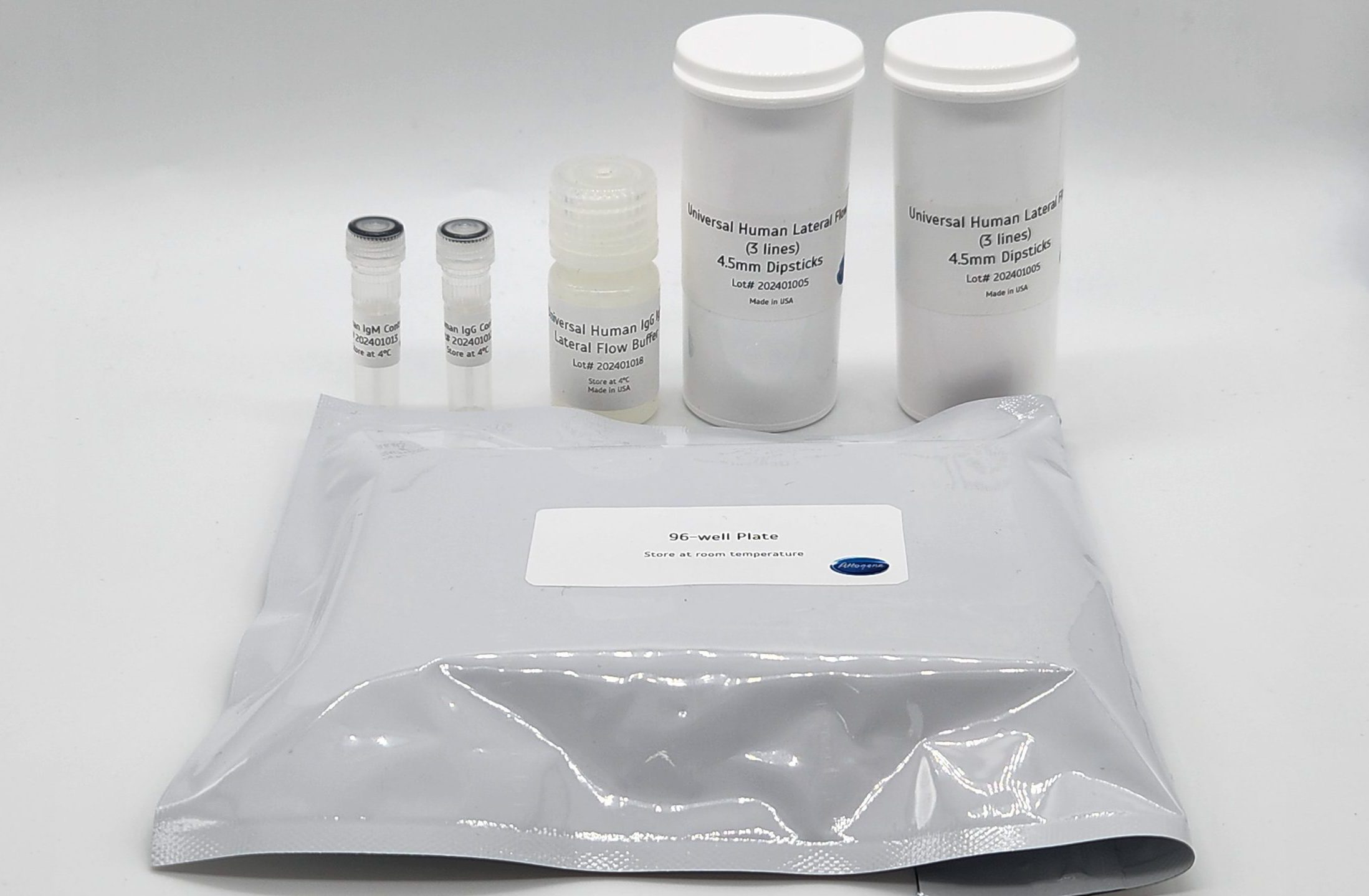
Fluorescent Universal Human IgG/IgM Lateral Flow Serology Kit (Quantum Dots)
Facebook
X
Pinterest
Email
Antibody tests are a method of choice to determine if a person has been exposed to a pathogen or not. They are also incredibly valuable in the detection of autoantibodies that can be found in human autoimmune disorders. In this test, a biotinylated antigen (User supplied) is mixed with a biotinylated rabbit IgG (bind to goat anti rabbit control line) and sample (human sera or plasma) is simply mixed into with the specially designed assay running buffer in a well of the supplied 96-well plate, mixed and is then added to the sample port of the cassette. Generally, the reaction is complete in 10-15 minutes. It is very important to note that the relative stoichiometry between the biotinylated antigen, biotinylated rabbit IgG added, and the streptavidin gold is critical for assay optimization. The appropriate concentration of biotinylated antigen to use with strips is dependent upon the purity and sequence and a standard curve can be used to determine the relative ratio (generally between 1ng-100ng per test). A positive control line (biotin-rabbit IgG) antibody will bind to the goat anti rabbit (GAR) line on the test to ensure the assay is running appropriately.
Attogene’s Human IgG/IgM universal fluorescent lateral flow assay kit is a ready-to-use, universal test strip, which is based on the lateral flow technology that uses 655nm Emission Quantum Dot particles containing streptavidin to conveniently capture biotinylated antigens. The device is designed to easily develop qualitative or quantitative rapid test systems for detection of anti-human IgG and IgM antibody that react to the any antigen that can be biotinylated (i.e. viral antigen, autoimmune antigen) and is easily customizable providing every laboratory with the possibility to perform assay feasibility.
Formats (fluorescent broad range UV light excitation range of 300nm to 400nm, 610nm emission) Streptavidin conjugate pad):
Other Products
[PM5000] ExcelBand™ 3-color Pre-Stained Protein Ladder, Regular Range (9-180 kDa), 250 μl x 2
Product Info
Document
Product Info
Description
The PM5000 ExcelBand™ 3-color Pre-Stained Protein Ladder Regular Range is a ready-to-use three-color protein standard with 13 pre-stained proteins covering a wide range of molecular weights from 10 to 180 kDa in Tris-Glycine Buffer (9 to 170 kDa in Bis-Tris (MOPS) buffer and 10 to 170 kDa Bis-Tris (MES) buffer). Proteins are covalently coupled with different chromophores for easy identification of bands, with three reference proteins carrying enhanced intensity corresponding to a blue band at 20 kDa, green at 40 kDa, and red at 75 kDa, respectively, as separated on SDS-PAGE (Tris-Glycine buffer). The PM5000 ExcelBand™ 3-color Pre-Stained Protein Ladder Regular Range is designed for monitoring protein separation during SDS-polyacrylamide gel electrophoresis, verification of Western transfer efficiency on membranes (PVDF, nylon, or nitrocellulose) and for approximating the size of proteins.
Features
Ready-to-use — Premixed with a loading buffer for direct loading, no need to boil.
Three reference bands — 75 kDa (red), 40 kDa (green), and 20 kDa (blue)
Contents
Approximately 0.1~0.4 mg/ml of each protein in the buffer (20 mM Tris-phosphate (pH 7.5 at 25°C), 2% SDS, 0.2 mM DTT, 3.6 M urea, and 15% (v/v) glycerol).
Quality Control
Under suggested conditions, PM5000 ExcelBand™ 3-color Pre-Stained Protein Ladder Regular Range resolves 13 major bands in SDS-PAGE (Tris-Glycine, MOPS, and MES buffer) and after Western blotting to nitrocellulose membrane.
Storage
4°C for 3 months -20°C for long term storage
Document
The PM5000 ExcelBand™ 3-color Pre-Stained Protein Ladder Regular Range is a ready-to-use three-color protein standard with 13 pre-stained proteins covering a wide range of molecular weights from 10 to 180 kDa in Tris-Glycine Buffer (9 to 170 kDa in Bis-Tris (MOPS) buffer and 10 to 170 kDa Bis-Tris (MES) buffer). Proteins are covalently coupled with different chromophores for easy identification of bands, with three reference proteins carrying enhanced intensity corresponding to a blue band at 20 kDa, green at 40 kDa, and red at 75 kDa, respectively, as separated on SDS-PAGE (Tris-Glycine buffer). The PM5000 ExcelBand™ 3-color Pre-Stained Protein Ladder Regular Range is designed for monitoring protein separation during SDS-polyacrylamide gel electrophoresis, verification of Western transfer efficiency on membranes (PVDF, nylon, or nitrocellulose) and for approximating the size of proteins.
Free-circulating nucleic acids, such as tumor-specific extracellular DNA fragments and mRNAs in the blood or fetal nucleic acids in maternal blood, are present in serum or plasma usually as short fragments, <1000bp (DNA). HiPure Circulating DNA Mini Kit enables efficient purification of these circulating nucleic acids from human plasma, serum, or urine. Samples can be fresh or frozen (provided that they have not been frozen and thawed more than once).
Details
Specifications
Features
Specifications
Main Functions
Isolation circulating DNA from 1ml plasma, serum, body fluids
Applications
qPCR, liquid or solid chip analysis, hybridization and SNP detection, etc.
Purification method
Mini spin column
Purification technology
Silica technology
Process method
Manual (centrifugation or vacuum)
Sample type
Serum, plasma and other cell-free fluid samples
Sample amount
1ml
Elution volume
≥30μl
Time per run
≤50 minutes
Liquid carrying volume per column
800µl
Binding yield of column
100µg
Principle
This product is based on silica gel purification. The sample is lysed and digested with lysate and protease, DNA is released into the lysate. Transfer to an adsorption plate and filter column. Nucleic acid is adsorbed on the membrane, while protein is not adsorbed and is removed with filtration. After washing proteins and other impurities, nucleic acid was finally eluted with low-salt buffer (10 Mm Tris,pH 8.0).
Advantages
High yield – most optimal process, free DNA (>50bp) can be obtained to the maximum extent
High concentration – low elution volume, ensuring high nucleic acid concentration
High purity – low alcohol binding method, completely removing inhibitor and protein pollution
High recovery – DNA can be recovered at the level of PG by silica gel column purification
Kit Contents
Contents
D318102
D318103
Purification Times
50 Preps
250 Preps
Buffer ACL
50 ml
250 ml
Buffer ACB*
60 ml
300 ml
Buffer DCW1*
22 ml
88 ml
Buffer DCW2*
10 ml
50 ml
Proteinase K
120 mg
540 mg
Protease Dissolve Buffer
10 ml
30 ml
Carrier RNA
110 μg
310 μg
Nuclease Free Water
10 ml
30 ml
HiPure CFDNA Mini Columns
50
250
2 ml Collection Tubes
100
500
Storage and Stability
Proteinase K and carrier RNA should be stored at 2–8°C upon arrival. However, short-term storage(up to 12 weeks) at room temperature (15–25°C) does not affect their performance. The remainingkit components can be stored dry at room temperature (15–25°C) and are stable for at least 18 months under these conditions.The entire kit can be stored at 2–8°C, but in this case buffers shouldbe redissolved before use. Make sure that all buffers are at room temperature when used.
Document
Free-circulating nucleic acids, such as tumor-specific extracellular DNA fragments and mRNAs in the blood or fetal nucleic acids in maternal blood, are present in serum or plasma usually as short fragments,