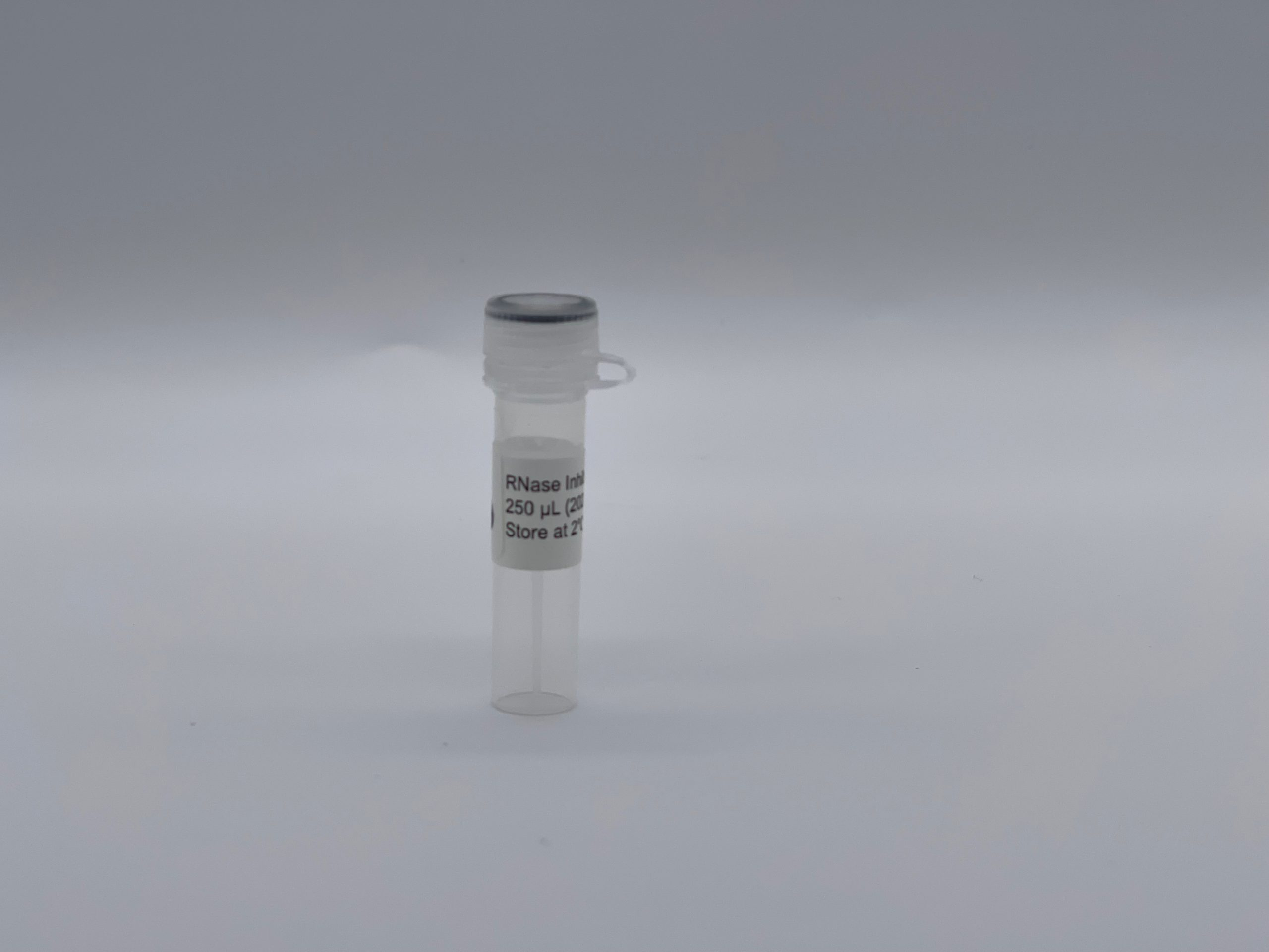
RNase Inhibitor (10,000 units) Concentration: 40 units/μL Volume: 0.25 mL Storage Conditions: Store at 2°C – 8°C Inhibits: RNases A, B, and C Supplied in: 1X PBS with 0.05% Sodium Azide.
Attogene’ s RNase Inhibitor is a protein-based ribonuclease inhibitor which noncovalently binds and inactivates a wide variety of RNases in a range of temperature (37–65°C) and pH (5.5–8.5) conditions. This product is distinct from placental RNase Inhibitor protein in that it inactivates RNases I and T1 in addition to RNases A, B and C. NA2021 is distinct from RNase Inhibitor protein in that it is less expensive, have more robust interactions with RNAses.
MADE AND FUNCTIONALLY TESTED AT ATTOGENE (MADE IN AUSTIN TEXAS – USA)
Other Products
Cerillo Stratus Plate Reader (600nm)
Product Info
Document
Product Info
Cerillo’s Stratus Plate Reader is a third party module that can be placed next to or inside the OT-2 or Opentrons Flex for post experimental analysis. The worktable adapter will allow you to utuilize the OT-2 or Opentrons Flex to prepare the samples directly into the plate reader positions on the worktable.
This small portable device is primarily for microbial growth curve analysis. The Stratus is compatible with Cerillo’s Canopy wireless device that allows real time visualization, control, and analytics. This product is compatible with reading 600nm wavelengths.
Document
Cerillo’s Stratus Plate Reader is a third party module that can be placed next to or inside the OT-2 or Opentrons Flex for post experimental analysis. The worktable adapter will allow you to utuilize the OT-2 or Opentrons Flex to prepare the samples directly into the plate reader positions on the worktable.
This small portable device is primarily for microbial growth curve analysis. The Stratus is compatible with Cerillo’s Canopy wireless device that allows real time visualization, control, and analytics. This product is compatible with reading 600nm wavelengths.
Magen’s HiPure columns are prepared by high quality glass fiber filter membrane as raw materials through membrane cutting, membrane release, ring release, ring pressing, gland, weighing and other processes. HiPure nucleic acid adsorption columns have the characteristics of long-term stability and high binding capacity. Experiments show that the highest binding capacity and binding efficiency of HiPure nucleic acid adsorption columns are basically unchanged when stored at room temperature for 4 years.
The series of nucleic acid columns produced by Magen Biotech are based on carefully selected imported glass fiber membranes (GF/B, GF/D, GF/F). Columns production processes such as polypropylene injection molding materials, injection molding process, and downstream membrane packing and compression rings are strictly controlled. This is to ensure that the column has extremely high adsorption capacity and long-term stability. Compared with conventional products on the market, Magen’s columns are with varieties, and binding rate will not change when stored at room temperature for 4 years.
Details
Specifications
Features
Specifications
Recommended application
Plasmid Maxi preparation
Preservation conditions
Room temperature
Stability
Up to 4 years
Filter membrane
High quality glass fiber filter GF/B, 8 layers
Membrane aperture
1.0 μm
Maximum binding yield of plasmid
1 mg
Maximum yield of alcohol mediated binding
5 mg
Plasmid yield
Up to 1mg
Single liquid carrying capacity of column
20 ml
Minimum elution volume
800 μl
Withstand centrifugal force
3,000-5,000 x g
Centrifuge
Low speed centrifuge for 50ml centrifuge tubes, >3000 x g, swing-out Rotor
Adsorption Mechanism
Based on the negatively charged DNA skeleton, it has a high affinity for positively charged glass fibers. In high salt and ethanol solutions, DNA/RNA binds to glass fiber and interacts with hydrophilic matrix on silica through hydrogen bond. DNA/RNA is tightly bound. All pollutants can be removed by washing solution. At high salt concentration, nucleic acids selectively bind to silicagel membrane, while other pollutants, mainly proteins, are removed by membrane washing.
Ordering information
CAT.No.
Product Name
Package
C13123
HiPure DNA Maxi Column III (8 x GF/B)with 50ml Collection Tubes
100/Bag
Purchase Guide
Item No.
Product Name
Membrane type/number of layers
Collection tubes
Plasmid DNA binding capacity (Physical adsorption)
Note: GF/B pore size is for 1.0μM glass fiber membrane; GF/F pore size is for 0.7μm glass fiber membrane.
Document
Magen’s HiPure columns are prepared by high quality glass fiber filter membrane as raw materials through membrane cutting, membrane release, ring release, ring pressing, gland, weighing and other processes. HiPure nucleic acid adsorption columns have the characteristics of long-term stability and high binding capacity. Experiments show that the highest binding capacity and binding efficiency of HiPure nucleic acid adsorption columns are basically unchanged when stored at room temperature for 4 years.
Food and beverage samples such as milk or milk based products, wine, fruit juices, paper (and cardboard), waste treatment samples and other materials (e.g. biological cultures etc.).
This product has been discontinued.
The hydrogen peroxide Assay Kit provides a simple robust method for the measurement of hydrogen peroxide. This kit utilizes a highly sensitive Megaplex Red probe which in the presence of horseradish peroxidase (HRP), allows for the measurement of H2O2 in a sample. Upon oxidation of the probe by HRP, a coloured product (resorufin) is formed that can be measured in fluorescence mode using excitation in the range 530-560 nm and emission at ~ 590 nm or colourimetrically at 570 nm. The excitation and emission maxima of Megaplex Red are 571 nm and 585 nm respectively.
Fluorometric/UV method for the determination of H2O2 content in a variety of samples.
Advantages
Simple format
Very competitive price (cost per test)
Ability to run in fluorescence or absorbance mode
User friendly – Detailed protocol provided for the creation of calibration curve and the calculation of concentration in fluorescence mode. Single point standard for quicker and simpler analysis in absorbance mode.
Mega-Calc™ software tool is available from our website for hassle-free raw data processing
Suitable for manual, microplate and auto-analyser formats
All reagents stable for > 2 years
Document
The hydrogen peroxide Assay Kit provides a simple robust method for the measurement of hydrogen peroxide. This kit utilizes a highly sensitive Megaplex Red probe which in the presence of horseradish peroxidase (HRP), allows for the measurement of H2O2 in a sample. Upon oxidation of the probe by HRP, a coloured product (resorufin) is formed that can be measured in fluorescence mode using excitation in the range 530-560 nm and emission at ~ 590 nm or colourimetrically at 570 nm. The excitation and emission maxima of Megaplex Red are 571 nm and 585 nm respectively.