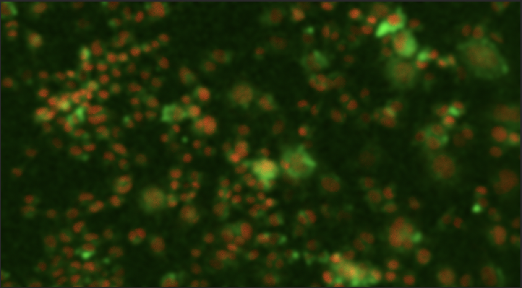
NeuroTarget Transfection Agent which is composed of a cation tagged rabies viral glycoprotein (RVG). Experimentally shown to deliver miRNA inside neuronal cell lines containing the nicotinic acetylcholine receptor (nAChR). Highly efficient delivery for the modulation of gene expression and cellular engineering.
Formulation of the rabies viral glycoprotein containing a positively charged tail used for association with nucleic acids. This protein enables the selective delivery of the nucleic acid into neuronal cells.
This peptide is a 29 amino acid fragment derived from rabies virus glycoprotein (RVG). Because neurotropic viruses cross the blood-brain barrier to infect brain cells, the same strategy may be used to enter the central nervous system and deliver siRNA to the brain. This peptide specifically binds to the acetylcholine receptor expressed by neuronal cells.
• NeuroTarget Transfection Agent
Other Products
Large Capacity High Speed Refrigerated Centrifuge
Product Info
Document
Product Info
GL12 Technical Parameter:
Max. Speed
12000rpm
Max. RCF
22650×g
Max. Capacity
4×1000ml
Timer
1~9h59min
RPM/RCF Convert
Yes
Noise (dB)
≤ 60
Temperature Range
-20~40℃
Acc/Dec
10 Kinds
Speed Accuracy
±20r/min
Temperature Accuracy
±1℃
Voltage(V/Hz)
AC 220V/110V 50HZ/60HZ
Size (L x W x Hmm)
780×675×830mm
Net Weight(Kg)
260KG
Certificates
CE,ISO & Calibration report are available
Matched Rotors for GL12
Order No.
Rotor No.
Max Speed (rpm)
Max Volume(ml)
Max.RCF(×g)
L12-1
Angle Rotor
12000
6×250/300ml
22650
L12-2
Angle Rotor
8000
6×500ml
11740
L12-3
Swing Rotor
4000
4×1000ml
4060
L12-4
Microplate Rotor
4000
4×4×96well
2940
L12-5
4000
2×4×96well
2490
L12-6
Angle Rotor
10000
4×500ml
16110
Document
Features
1. Widely used in the field of biochemistry, biological products, pharmaceutical factory and laboratory.
2. Brushless frequency motor for model GL12 which in great torque, free maintenance, no powder pollution, quick in speed up and down.
3. Model GL12 with the digital display which indicates the speed、time and temperature.
4. Micro computer control, there are 10 kinds of program and 10 kinds of acceleration and deceleration for your choice.
5. There are high speed rotors and low speed rotors for your choice.
6. Electric lid lock, compact design, super speed and imbalance protection.
7. The centrifuge body is made of high-quality steel, safe and reliable.
resDNASEQ E.coli Residual DNA Quantitation kit (Without DNA Control)
Product Info
Document
Product Info
Description
Features of the sDNASEQ E.coli Residual DNA
Quantitation kit include:
Simpler and Rapid
Only three steps will be need for Sample Preparation and All components of the Sample Preparation Kit can be stored at room temperature.
Only one Reagent for qPCR;
Only 1.5 hours will be needed for the whole test.
Accurate
Perfect amplification curve, good amplification efficiency and good precision.
Highly sensitive quantitation using proven TaqMan™ real-time qPCR technology.
Limit of Detection (LOD): 1 pg/mL; Limit of Quantification (LOQ): 5 pg/mL
The recovery rate of different concentration samples in the linear range is between 70% and 130%
Kit Performance
Fig 1. Only three steps will be need for Sample Preparation and only 20 minitutes will be taken for Sample Preparation.
Fig 2. Seven concentration samples of 5fg/μL, 10fg/μL, 20fg/μL, 30fg/μL, 300fg/μL, 3pg/μL, 30pg/μL, 300pg/μL were detected. CV of each concentration was < 30%, Regression coefficient associated with standard solutions was 0.99975, and amplification efficiency was 100.068%.
Fig 3. Four concentration samples of 5fg/μL, 10fg/μL, 20fg/μL, and 30fg/μL were detected, and 10 multiple holes were detected for each concentration. The detection values of 5fg/μL and above were CV <30%.
Fig 4. DNA recovery can be determined by including samples spiked with known DNA amounts which are prepared from the corresponding DNA standards. Typically, the range for this value varies from 70% to 130%.
Fig 5. Only one Reagent for qPCR MIX.
Document
Note: Price not include shipment & duty, contact us to get full quote. The resDNASEQ E.coli Residual DNA Quantitation kit is designed for the quantification of residual DNA from E. coli, in cell lines which are used for production of biopharmaceutical products. The resDNASEQ E.coli Residual DNA Quantitation kit use TaqManTM quantitative PCR to perform rapid, specifc quantitation of femtogram levels of residual host-cell or plasmid DNA. The kit was developed to meet the sensitivity requirements defined by WHO (10 ng E. coli DNA per therapeutic dose).
[PS1000] FluoroStain™ Protein Fluorescent Staining Dye (Red, 1,000X), 1 ml
Product Info
Document
Product Info
Description
The FluoroStain™ Protein Fluorescent Staining Dye (Red, 1000×) is designed to substitute the common Coomassie Blue protein staining method, offering greater sensitivity and ease of operation. Unlike Coomassie Blue stain, the FluoroStain Protein Fluorescent Staining Dye binds to protein with high specificity, making destaining process an option rather than a requirement. With further reduction of background signals via destaining process, the FluoroStain™ is capable of achieving detection level parallel to silver staining without specialized imaging equipment, making it one of the most sensitive dyes available. In addition to its remarkable sensitivity, the FluoroStain™ Protein Fluorescent Staining Dye (Red, 1000×) brings a more reliable and safer user experience, since the stained gel can be visualized with blue-light illumination, avoiding the risk of skin/ eye damage caused by UV light. For best result, we suggest using B-BOX™ Blue Light LED epi-illuminator to visualize and analyze the gel stained with FluoroStain Protein Fluorescent Staining Dye (Red, 1000×). The FluoroStain™ Protein Fluorescent Staining Dye is compatible to the analysis of mass spectra, i.e. LC-MS/MS, MALDI-TOF, etc.
Spectral Characteristics
When it is bound with bovine serum albumin (BSA), the fluorescent emission of FluoroStain Protein Fluorescent Staining Dye can be excited by UV and blue light sources, with excitation peaks around 369 and 517 nm and emission at 605 nm. In absence of BSA, FluoroStain Protein Fluorescent Staining Dye shows ignorable fluorescence as compared with protein-bound form, therefore giving a clear background for photographic analysis.
These spectral characteristics made this fluorescent dye compatible with a wide variety of gel reading facilities, including UV/ blue light epi- and transilluminator, argon laser and mercury-arc lamp excitation gel scanners.
Storage
Protected from light -20°C for 24 months
Document
The FluoroStain™ Protein Fluorescent Staining Dye (Red, 1000×) is designed to substitute the common Coomassie Blue protein staining method, offering greater sensitivity and ease of operation. Unlike Coomassie Blue stain, the FluoroStain Protein Fluorescent Staining Dye binds to protein with high specificity, making destaining process an option rather than a requirement. With further reduction of background signals via destaining process, the FluoroStain™ is capable of achieving detection level parallel to silver staining without specialized imaging equipment, making it one of the most sensitive dyes available. In addition to its remarkable sensitivity, the FluoroStain™ Protein Fluorescent Staining Dye (Red, 1000×) brings a more reliable and safer user experience, since the stained gel can be visualized with blue-light illumination, avoiding the risk of skin/ eye damage caused by UV light. For best result, we suggest using B-BOX™ Blue Light LED epi-illuminator to visualize and analyze the gel stained with FluoroStain Protein Fluorescent Staining Dye (Red, 1000×). The FluoroStain™ Protein Fluorescent Staining Dye is compatible to the analysis of mass spectra, i.e. LC-MS/MS, MALDI-TOF, etc.