
Description
Specifications:
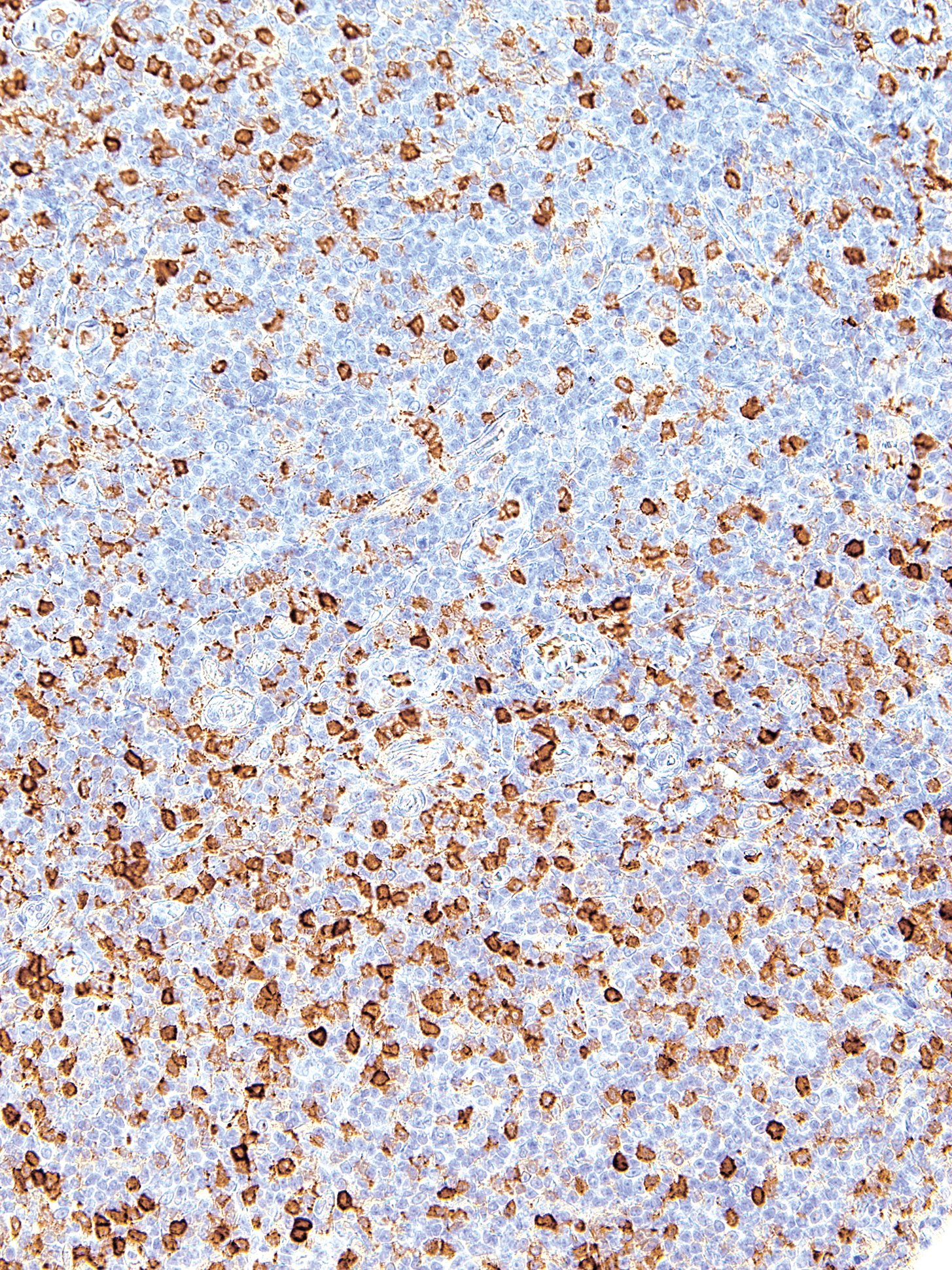
| Clone | IHC512 |
| Source | Mouse Monoclonal |
| Positive Control | Hairy Cell Leukemia |
| Dilution Range | 1:200 |

| Clone | IHC512 |
| Source | Mouse Monoclonal |
| Positive Control | Hairy Cell Leukemia |
| Dilution Range | 1:200 |

The MagPure Plasmid purification system uses the paramagnetic bead technology for high-throughput preparation of high-copy or low-copy plasmid DNA from E. coli cells. This kit also can be used with fosmid and BAC vector-based constructs. The system uses alkaline lysis followed by a MagPure purification to differentially bind plasmid DNA to paramagnetic beads. While the DNA is bound to the beads, contaminants can be rinsed away using a simple washing procedure. Because MagPure uses magnetic separation technology, the protocol does not require vacuum filtration. This makes kit extremely amenable to automation. Plasmid DNA purified with this system is most commonly used in Sanger Sequencing and PCR amplification.
Specifications
| Features | Specifications |
| Main Functions | Isolation up to 20µg plasmid DNA from 1-3ml bacterial culture |
| Applications | Enzyme digestion, sequencing, PCR and labeling, etc. |
| Purification technology | Magnetic beads technology |
| Process method | Manual or automatic |
| Sample type | Conventional plasmid, plasmid≤30KB |
| Sample amount | 1-3ml |
| Elution volume | ≥50μl |
This product is based on the purification method of high binding magnetic particles. The sample is lysed and digested under the action of lysate and Lysozyme. DNA is released into the lysate. After adding magnetic particles and binding solution, DNA will be adsorbed on the surface of magnetic particles, and impurities such as proteins will be removed without adsorption.The adsorbed particles were washed with washing solution to remove proteins and impurities, washed with ethanol to remove salts, and finally DNA was eluted by Elution Buffer.
Advantages
1. Suitable for extracting plasmids from 1-5ml or <3ml YT medium.
2. The same amount of buffer 1, 2, and 3 avoids errors caused by adjusting the pipette, making it convenient to use in conjunction with automated workstations.
3. Containing buffer 1 for washing, reducing the problem of false high production.
4. The purified plasmid can be directly used for sequencing, enzyme digestion, PCR, and other applications.
Kit Contents
| Contents | P181102 | P181103 | P181104 |
| Purification Times | 100 Preps | 500 Preps | 5000 Preps |
| RNase A | 10 mg | 50 mg | 2 x 250 mg |
| Buffer P1 | 30 ml | 150 ml | 2 x 750 ml |
| Buffer P2 | 30 ml | 150 ml | 2 x 750 ml |
| Buffer N3 | 30 ml | 150 ml | 2 x 750 ml |
| Buffer PW1 | 35 ml | 180 ml | 2 x 900 ml |
| MagPure Particle NB* | 2.2 ml | 11 ml | 2 x 60 ml |
Storage and Stability
RNase A and MagPure Particle NB should be stored at 2-8°C upon arrival. However, short-term storage (up to 24 weeks) at room temperature (15-25°C) does not affect its performance. The remaining kit components can be stored dry at room temperature (15-25°C) and are stable for at least 18 months under these conditions. After addition of RNase A, Buffer P1 is stable for 6 months when stored at 2-8°C.
Experiment Data

The MagPure Plasmid purification system uses the paramagnetic bead technology for high-throughput preparation of high-copy or low-copy plasmid DNA from E. coli cells. This kit also can be used with fosmid and BAC vector-based constructs. The system uses alkaline lysis followed by a MagPure purification to differentially bind plasmid DNA to paramagnetic beads. While the DNA is bound to the beads, contaminants can be rinsed away using a simple washing procedure. Because MagPure uses magnetic separation technology, the protocol does not require vacuum filtration. This makes kit extremely amenable to automation. Plasmid DNA purified with this system is most commonly used in Sanger Sequencing and PCR amplification.

Thin polyester heat sealing film which is easily pierceable with autosampler needles/ABI® 3730. The seal is suitable for PCR, qPCR and optical applications.
