
Description
Specifications
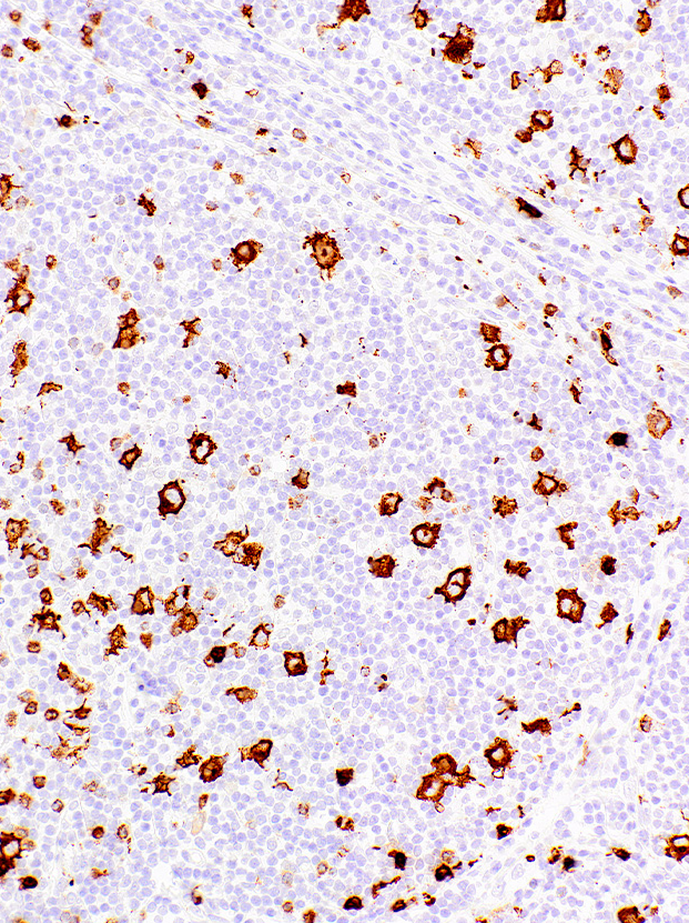
| Clone | IHC527 |
| Source | Mouse Monoclonal |
| Positive Control | Hodgkin’s Lymphoma |
| Dilution Range | 1:200 |

| Clone | IHC527 |
| Source | Mouse Monoclonal |
| Positive Control | Hodgkin’s Lymphoma |
| Dilution Range | 1:200 |

Attogene’s Microcystin Test Kit (Rapid – Recreational Water) can be used to detect microcystins in liquid samples; highly sensitive, rapid, robust screening kit for algae toxins microcystins and nodularins.
The most frequently reported cyanobacterial toxins are the hepatotoxic microcystins (MCs). MCs are peptides with a molecular weight ranging from 900 to 1,100 Da. They consist of seven amino acids of which the two terminal amino acids of the linear peptide are condensed to form a cyclic compound.
A tiered notification system which takes different actions based on different numeric thresholds for microcytin-LR concentrations in recreational waters has been developed. This is guidance that allows states to take various actions—such as posting information about harmful algal blooms (HABs), issuing a recreational public health advisory, or temporarily closing recreational waters through a no contact advisory—depending on the severity of the bloom event.
Microcystin-LR
Informational sign postings about HABs at recreational waters: < 6 μg/L
Recreational public health advisory: 6 μg/L
Elevated recreational public health advisory (e.g. no contact): 20 μg/L
Screening of Microcystins in water samples at 6 ppb or lower
Format: 10 tests (5 tests/5 control)
Water Sample Bottles
Negative Control
1mL Syringe
Sample Dilution Buffer also sold separately
Sample Filters

Biocolor’s Purple-Jelley assay kit is the perfect tool for accurate measurement of hyaluronic acid / Hyaluronan levels in your samples. This colorimetric assay is optimised for quantitative analysis in-vivo, tissue-derived hyaluronic acid / Hyaluronan and includes full step-by-step instructions.
Colorimetric Detection (655nm) (Endpoint)
Hyaluronic acid, in its hydrated form, is a unique carbohydrate polymer, often referred to as a ‘gentle giant.’ It consists of a lengthy, flexible, non-branching chain with a repeating disaccharide pattern. This disaccharide is composed of alternating uronic acid and aminosugar units.
Discovering the J-Aggregate Effect in Cyanine DyesIn 1936, Edwin Jelley made a fascinating observation, documented it in a letter to Nature (Nature 138, 1009 – 1010). He noted a peculiar behaviour of certain cyanine dyes, that when dissolved in 5 M NaCl, they dyes exhibited a third absorbance peak at a longer wavelength, around 650nm. In deionized water, however, they displayed only a double peak at approximately 540nm and 570nm. The 650nm peak in concentrated dye solutions resulted from the aggregation of dye molecules and was later termed a ‘J-aggregate,’ in honor of Edwin Jelley. The J-aggregate is known as a supra-molecular complex, formed by stacking individual dye molecules.
Subsequent research in the 1960s, notably by Kay et al. (J. Physical Chem. 68, 1896 – 1906), revealed that various biological polymers, including proteins, DNA, polar lipids, and glycosaminoglycans, could also induce this third absorbance peak. This phenomenon led to the development of the Purple-Jelley assay, named after the purple color of the dye reagent and Edwin Jelley himself.
During the assay, hyaluronic acid is selectively purified during the assay sample preparation protocol. This is then reacted with the Purple-Jelley dye reagent, and the absorption of the characteristic third wavelength recorded. By comparison with a calibration curve the hyaluronic acid content of the sample can be measured.
Step 1. The assay protocol takes tissue samples through a sequential sample preparation protocol which involves enzymatic protein digestion, followed by precipitation and purification of GAGs, culminating in the precipitation of purified Hyaluronic acid.
Step2. The processed sample is then incubated for 10 minutes with the Purple-Jelley dye reagent, forming a coloured product which can be measured spectrophotometrically.
Step 3. The Hyaluronic acid content of unknown samples can be calculated by comparison against a calibration curve prepared using a standard comprising hyaluronic acid (supplied with the kit).
10 – 100µg/ml
10µg/ml
Colorimetric Detection (655nm) (Endpoint)
100 in total (allows a maximum of 46 samples to be run in duplicate alongside a standard curve).
In-vivo: Hyaluronic acid purified from in-vivo tissues. The kit protocol involves extraction and purification of hyaluronic acid prior to reaction with the Purple-Dye reagent.
This kit is designed for research use only. Not for use in diagnostic procedures.
Kit requires access to a centrifuge, as well as a spectrophotometer/colorimeter capable of colorimetric, absorbance detection at 655nm.
Specific sample preparation protocols may require customer to provide further reagents, consult assay manual for further information.
Mode of ActionAssay SpecificationsKit Contents
1. Purple-Jelley Dye Reagent (1x 20ml)
2. Hyaluronan Reference Standard (1x 5ml, 0.2mg/ml soluble Hyaluronic Acid)
3. Precipitating Reagent (2x 34ml)
4. Sodium Chloride (1x 20ml)
5. Cetylpyridinium Chloride (1x 20ml)
6. TRIS-buffered Saline (5x tablets)
7. 2ml screw-cap tubes for preparation of samples.
8. Assay kit manual
NB: Additional reagents may be required for sample preparation prior to assay. Consult manual or contact us for further details.
Biocolor’s Purple-Jelley assay kit is the perfect tool for accurate measurement of hyaluronic acid / Hyaluronan levels in your samples. This colorimetric assay is optimised for quantitative analysis in-vivo, tissue-derived hyaluronic acid / Hyaluronan and includes full step-by-step instructions.

Biotin-PEG4-C1-alkyne is azide reactive biotinylation reagent, PEG4 arm increase aqueous solubility of the reagent. Reagent grade, for research purpose. Please contact us for GMP-grade inquiries.

Biotin-PEG4-C1-alkyne is azide reactive biotinylation reagent, PEG4 arm increase aqueous solubility of the reagent. Reagent grade, for research purpose. Please contact us for GMP-grade inquiries.