Usages: For cultivating of bacteria.And sterility test of drugs and biological products.
Principle: Tryptone, peptone and yeast extract multivalent powder provides a nitrogen source, vitamins, and growth factors; sodium chloride to maintain osmotic balance; glucose carbon source; dipotassium hydrogen phosphate as a buffering agent.
Formulation(per liter): Pancreatic Digest of Casein 17g Papaic Digest of Soybean 3g Sodium chloride 5g Dipotassium hydrogen phosphate 2.5g Glucose Monohydrate 2.5g Final pH 7.3±0.2
How to use: 1.Suspend 30g in 1L of distilled water , stirring heated to boiling to completely dissolve ,autoclave at 121℃ for 15 minutes. 2.Diluted and treated samples.
Storage: Keep container tightly closed, store in a cool, dry place, away from bright light. Storage period of 3 years.
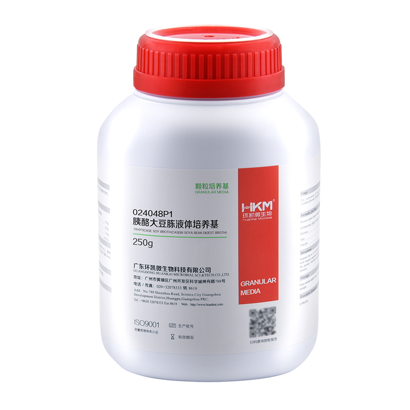
Our SNPsig® kits use our own proprietary genotyping method to enable the identification of SARS-CoV-2 variants of concern. These products can be used on any real-time PCR machine using familiar protocols, whilst resulting in exceptional genotyping data.
Positive control templates for wild-type and variants are supplied in every kit to make data interpretation simple.
Our SNPsig® technology provides an alternative to sequencing as well as S gene target failure (SGTF) that enables scientists to analyse and monitor these specific genomic mutations. Our kits can provide a pivotal role in screening for SARS-CoV-2 variants for the purpose of genomic surveillance and studies.
Document
For the detection of the SARS-CoV-2 variants with the N501Y mutation (UK, South Africa and Brazil)
Rapid detection of specific detection profiles
High priming efficiency
Sensitive to < 100 copies of target
Positive copy number standard curve for quantification
Accurate controls to confirm findings
96 reactions, includes master mix
This product is suitable for rapid extraction of DNA from tissue, cells, blood, saliva, swabs, blood spots, semen and other clinical samples. DNA can be used directly for PCR, quantitative PCR, Southern Blot, test of virus DNA and so on.
Details
Specifications
Features
Specifications
Main Functions
Isolation total DNA from 1-100ul blood, FFPE, tissue and other samples
Applications
Second generation sequencing, PCR, real timePCR, etc.
Purification method
Monodisperse magnetic beads
Purification technology
Magnetic beads technology
Process method
Manual or automatic
Sample type
Anticoagulant blood, concentrated blood, buffy coat, lymphocytes and cultured cells
Sample amount
Body fluid : 10 – 200μl, Tissue : ≤10mg
Yield
10ng – 15μg
Elution volume
80 -100μl
Time per run
Principle
This product is based on the purification method of high binding magnetic particles. The sample is lysed and digested under the action of lysate and Protease. DNA is released into the lysate. After adding magnetic particles and binding solution, DNA will be adsorbed on the surface of magnetic particles, and impurities such as proteins will be removed without adsorption. The adsorbed particles were washed with washing solution to remove proteins and impurities, washed with ethanol to remove salts, and finally DNA was eluted by Elution Buffer.
Advantages
High binding force – suitable for handling DNA rich samples, such as whole blood, buffy coat, concentrated blood, etc.
High purity – carboxyl magnetic beads with weak surface adsorption, getting higher purity
General – can be used for various clinical samples
Kit Contents
Contents
IVD3101
Purification Times
200
MagBind Particles
4.5 ml
Proteinase K
90 mg
Protease Dissolve Buffer
10 ml
Buffer ATL
60 ml
Buffer AL
60 ml
Buffer BD*
20 ml
Buffer BXW1*
110 ml
Elution Buffer
30 ml
Storage and Stability
Proteinase K, MagBind Particles should be stored at 2–8°C upon arrival. However, short-termstorage (up to 18 weeks) at room temperature (15–25°C) does not affect their performance. The remainingkit components can be stored dry at room temperature (15–25°C) and are stable for at least 18 months under these conditions.
Document
This product is suitable for rapid extraction of DNA from tissue, cells, blood, saliva, swabs, blood spots, semen and other clinical samples. DNA can be used directly for PCR, quantitative PCR, Southern Blot, test of virus DNA and so on.
[PM2610] ExcelBand™ Enhanced 3-color High Range Protein Marker (9-245 kDa), 250 μl x 2
Product Info
Document
Product Info
Description
The PM2610/PM2611 ExcelBand™ Enhanced 3-color High Range Protein Marker is a ready to use three-color protein standard with 12 pre-stained proteins covering a wide range of molecular weights from 10 to 245 kDa in Tris-Glycine buffer (9 to 235 kDa in BisTris (MOPS) buffer and 10-235 kDa in Bis-Tris (MES) buffer). Proteins are covalently coupled with different chromophores for easy identification of bands, with two reference proteins carrying enhanced intensity corresponding to a green at 25 kDa and red at 75 kDa, respectively, as separated on SDS-PAGE (Tris-Glycine buffer). The ExcelBand™ Enhanced 3-color High Range Protein Marker is designed for monitoring protein separation during SDS-polyacrylamide gel electrophoresis, verification of Western transfer efficiency on membranes (PVDF, nylon, or nitrocellulose) and for approximating the size of proteins.
Features
Ready-to-use — Premixed with a loading buffer for direct loading, no need to boil.
Two reference bands — 75 kDa (red) and 25 kDa (green)
Contents
Approximately 0.2~0.6 mg/ml of each protein in the buffer (20 mM Tris-phosphate (pH 7.5 at 25°C), 2 % SDS, 3.6 M Urea, and 15 % (v/v) Glycerol).
Quality Control
Under suggested conditions, the PM2610/PM2611 Enhanced 3-color High Range Protein Marker resolves 12 major bands in 15% SDS-PAGE (Tris-Glycine buffer, MOPS, and MES buffer) and after Western blotting to nitrocellulose membrane.
Storage
4°C for 3 months -20°C for long term storage
Document
The PM2610/PM2611 ExcelBand™ Enhanced 3-color High Range Protein Marker is a ready to use three-color protein standard with 12 pre-stained proteins covering a wide range of molecular weights from 10 to 245 kDa in Tris-Glycine buffer (9 to 235 kDa in BisTris (MOPS) buffer and 10-235 kDa in Bis-Tris (MES) buffer). Proteins are covalently coupled with different chromophores for easy identification of bands, with two reference proteins carrying enhanced intensity corresponding to a green at 25 kDa and red at 75 kDa, respectively, as separated on SDS-PAGE (Tris-Glycine buffer). The ExcelBand™ Enhanced 3-color High Range Protein Marker is designed for monitoring protein separation during SDS-polyacrylamide gel electrophoresis, verification of Western transfer efficiency on membranes (PVDF, nylon, or nitrocellulose) and for approximating the size of proteins.