
Introduction
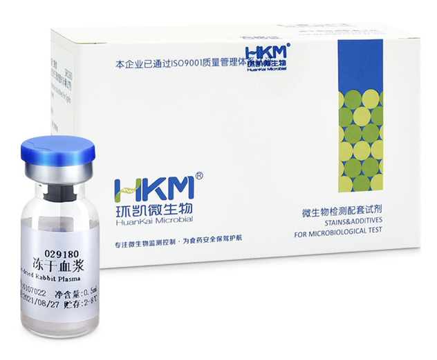
029180 Freeze-dried Rabbit Plasma
Usage:For coagulase test of Staphylococcus aureus.
Specification:0.5ml*10vials

029180 Freeze-dried Rabbit Plasma
Usage:For coagulase test of Staphylococcus aureus.
Specification:0.5ml*10vials

This product provides fast and easy methods for purification of total DNA for reliable PCR and Southern blotting. Total DNA(e.g., genomic, viral, mitochondrial) can be purified from small volume of blood, tissue and dry blood spots.
Specifications
| Features | Specifications |
| Main Functions | Isolation total DNA from 1-10μl blood, <10mg tissue, urine, blood stain, seminal stain |
| Applications | PCR, southern bolt and virus detection, etc. |
| Purification method | Mini spin column |
| Purification technology | Silica technology |
| Process method | Manual (centrifugation or vacuum) |
| Sample type | Animal tissues, blood stain, urine, seminal stain and various forensic samples |
| Sample amount | Blood:1-100μl, Tissue:<10mg |
| Elution volume | |
| Time per run | |
| Liquid carrying volume per column | |
| Binding yield of column |
This product is based on silica Column purification. The sample is lysed and digested with lysate and protease, DNA is released into the lysate. Transfer to an adsorption column. Nucleic acid is adsorbed on the membrane, while protein is not adsorbed and is removed with filtration. After washing proteins and other impurities, nucleic acid was finally eluted with low-salt buffer (10mmTris, pH9.0, 0.5mm EDTA).
Kit Contents
| Contents | D312502 | D312503 |
| Purification Times | 50 Preps | 250 Preps |
| Buffer ATL | 15 ml | 60 ml |
| Buffer AL | 15 ml | 60 ml |
| Buffer GW1* | 22 ml | 66 ml |
| Buffer GW2* | 20 ml | 2 x 50 ml |
| Carrier RNA | 310 μg | 2 x 310 µg |
| Proteinase K | 24 mg | 120 mg |
| Protease Dissolve Buffer | 1.8 ml | 10 ml |
| Buffer AE | 15 ml | 60 ml |
| HiPure DNA Mini Columns I | 50 | 2 x 125 |
| 2 ml Collection Tubes | 100 | 5 x 100 |
Storage and Stability
Carrier RNA and Proteinase K should be stored at 2–8°C upon arrival. However, short-term storage (up to 12 weeks) at room temperature (15–25°C) does not affect its performance. The remaining kit components can be stored dry at room temperature (15–25°C) and are stable for at least 18 months under theseconditions.
Experiment Data

This product provides fast and easy methods for purification of total DNA for reliable PCR and Southern blotting. Total DNA(e.g., genomic, viral, mitochondrial) can be purified from small volume of blood, tissue and dry blood spots.

Nucleic acid testing (NAT) is the method of choice for detection and quantification of a wide range of micro organisms. Primerdesign manufactures and supplies high quality quantitative real-time PCR kits for the detection and simultaneous quantification of numerous significant pathogens . A copy number standard curve is provided for quantification and an the internal extraction template (DNA or RNA), controls for the quality of the nucleic acid extraction and eliminates false negative results.
The kit is designed with the broadest possible detection profile to ensure that all clinically relevant strains and subtypes are detected. Target sequences are selected by working with data from key opinion leaders in the field. Multiple sequence alignments and unprecedented real-time PCR expertise in design and validation ensure the best possible kit.
Details of the target and priming specificity are included in the individual handbooks above.
Packaged, optimised and ready to use. Expect Better Data.
Exceptional value for money
Rapid detection of all clinically relevant subtypes
Positive copy number standard curve for quantification
Highly specific detection profile
High priming efficiency
Broad dynamic detection range (>6 logs)
Sensitive to < 100 copies of target
Accurate controls to confirm findings

Magen’s HiPure columns are prepared by high quality glass fiber filter membrane as raw materials through membrane cutting, membrane release, ring release, ring pressing, gland, weighing and other processes. HiPure nucleic acid adsorption columns have the characteristics of long-term stability and high binding capacity. Experiments show that the highest binding capacity and binding efficiency of HiPure nucleic acid adsorption columns are basically unchanged when stored at room temperature for 4 years.
The series of nucleic acid columns produced by Magen Biotech are based on carefully selected imported glass fiber membranes (GF/B, GF/D, GF/F). Columns production processes such as polypropylene injection molding materials, injection molding process, and downstream membrane packing and compression rings are strictly controlled. This is to ensure that the column has extremely high adsorption capacity and long-term stability. Compared with conventional products on the market, Magen’s columns are with varieties, and binding rate will not change when stored at room temperature for 4 years.
Specifications
| Features | Specifications |
| Recommended application | Small amounts of nucleic acid isolation, viral nucleicacid from cell free samples |
| Preservation conditions | Room temperature |
| Stability | Up to 4 years |
| Filter membrane | High quality glass fiber filter GF/F, 3 layers |
| Membrane aperture | 0.7μm |
| Maximum binding yield of plasmid | 30 μg |
| Maximum yield of alcohol mediated Binding | 200 μg |
| Single liquid carrying capacity of column | 800 μl |
| Minimum elution volume | 30 μl |
| Withstand centrifugal force | 16,000 x g |
| Centrifuge | Small high speed centrifuge (2ml) |
Adsorption Mechanism
Based on the negatively charged DNA skeleton, it has a high affinity for positively charged glass fibers. In high salt and ethanol solutions, DNA/RNA binds to glass fiber and interacts with hydrophilic matrix on silica through hydrogen bond. DNA/RNA is tightly bound. All pollutants can be removed by washing solution. At high salt concentration, nucleic acids selectively bind to silicagel membrane, while other pollutants, mainly proteins, are removed by membrane washing.
Ordering information
| CAT.No. | Product Name | Package |
| C13112 | HiPure Viral Mini Column I (3 x GF/F)with 2ml Collection Tubes | 1000/Bag |
| Item No. | Product Name | Membrane type/number of layers | Collection tubes | Plasmid DNA binding capacity (Physical adsorption) | gDNA/RNA binding capacity (Alcohol-mediated adsorption) | Minimum Elution volume | Liquid volume capacity |
| C13010 | HiPure DNA Nano Column | 2 layers GF/F | 2ml without cap | 5μg | 20μg | 10μl | 700μl |
| C13011 | HiPure DNA Micro Column | 3 layers GF/F | 2ml without cap | 10μg | 50μg | 15μl | 700μl |
| C13100 | HiPure DNA Mini Column I | 2 layers GF/B | 2ml without cap | 15μg | 100μg | 30μl | 700μl |
| C13110 | HiPure DNA Mini Column II | 4 layers GF/B | 2ml without cap | 35μg | 200μg | 50μl | 800μl |
| C13111 | HiPure RNA Mini Column | 3 layers GF/B | 2ml without cap | 30μg | 200μg | 30μl | 800μl |
| C13112 | HiPure Viral Mini Column | 3 layers GF/F | 2ml without cap | 30μg | 200μg | 30μl | 800μl |
| C13113 | HiPure CFDNA Mini Column | 3 layers GF/F,1 layer GF/B | 2ml without cap | 30μg | 200μg | 30μl | 800μl |
| C13120 | HiPure DNA Midi Column | 4 layers GF/B | 15ml Centrifuge tube | 125μg | 1mg | 500μl | 4ml |
| C13121 | HiPure DNA Midi Column III | 8 layers GF/B | 15ml Centrifuge tube | 250μg | 1mg | 500μl | 4ml |
| C13122 | HiPure DNA Maxi Column | 4 layers GF/B | 50ml Centrifuge tube | 500μg | 5mg | 1000μl | 20ml |
| C13123 | HiPure DNA Maxi Column III | 8 layers GF/B | 50ml Centrifuge tube | 1mg | 5mg | 1000μl | 20ml |
| C13124 | HiPure DNA Maxi Column C | 8 layers GF/B | 50ml high speed Centrifuge tube | 1mg | 5mg | 700μl | 12ml |
| C13130 | HiPure DNA Plate | 2 layers GF/F | 1.6ml Plate | 30μg | 100μg | 80μl | 900μl |
| C13131 | HiPure gDNA Plate | 2 layers GF/B | 1.6ml Plate | 30μg | 100μg | 80μl | 900μl |
Note: GF/B pore size is for 1.0μM glass fiber membrane; GF/F pore size is for 0.7μm glass fiber membrane.
Magen’s HiPure columns are prepared by high quality glass fiber filter membrane as raw materials through membrane cutting, membrane release, ring release, ring pressing, gland, weighing and other processes. HiPure nucleic acid adsorption columns have the characteristics of long-term stability and high binding capacity. Experiments show that the highest binding capacity and binding efficiency of HiPure nucleic acid adsorption columns are basically unchanged when stored at room temperature for 4 years.