


Opentrons OT-2 Single-Channel Electronic Pipettes automate liquid handling and pipetting for most daily wetlab applications. Best for low to medium throughput workflows with high precision.
Opentrons OT-2 Single-Channel Electronic Pipettes automate liquid handling and pipetting for most daily wetlab applications. Best for low to medium throughput workflows with high precision.

DBCO-PEG8-NHS Ester is a click chemistry molecule cosisting of an NHS ester that is reactive specifically and efficiently with primary amines (e.g. the side chain of lysine residues or aminosilane-coated surfaces) at neutral or slightly basic condition to form a covalent bond. PEG8 spacer arm improves water solubility and provides a long and flexible connection that minimizes steric hindrance involved with ligation. DBCO is commonly used for copper-free Click Chemistry reactions. Reagent grade, for research purpose. Please contact us for GMP-grade inquiries.

DBCO-PEG8-NHS Ester is a click chemistry molecule cosisting of an NHS ester that is reactive specifically and efficiently with primary amines (e.g. the side chain of lysine residues or aminosilane-coated surfaces) at neutral or slightly basic condition to form a covalent bond. PEG8 spacer arm improves water solubility and provides a long and flexible connection that minimizes steric hindrance involved with ligation. DBCO is commonly used for copper-free Click Chemistry reactions. Reagent grade, for research purpose. Please contact us for GMP-grade inquiries.

Over the past several decades, mitochondrial DNA (mtDNA) has played an increasingly important role in forensic analyses of various criminal cases. A few hairs left at a crime scene contain enough mtDNA for extraction. The hair shaft, which protrudes out of the scalp, does not contain any nuclear DNA. It does, however, contain mtDNA. While nuclear DNA is present in only two copies per cell, the small circular mtDNA molecule is present in hundreds to thousands of copies per cell making it very abundant. Mitochondrial DNA is maternally inherited, and all of a woman’s offspring will have the same mtDNA profile. An advantage of this is that a single maternal relative of that person may provide a reference sample for comparison to a sample found at a crime scene.
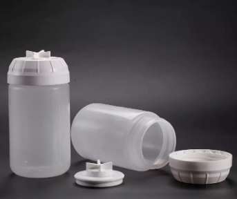
Norgen’s Hair Mitochondrial DNA Isolation Kit provides a fast, reliable, and simple procedure for isolating mtDNA from hair shafts. Purification is based on spin column chromatography and the DNA is preferentially purified from other components. Typical yields will vary depending on the sample input volume used. The purified DNA is compatible with all downstream applications including PCR and NGS.
Figure 1 / 4

Click for expanded view
Storage Conditions and Product Stability
Store DTT at -20°C upon arrival. All other solutions should be kept tightly sealed and stored at room temperature. These reagents should remain stable for at least 1 year in their unopened containers. The kit contains a ready to-use Proteinase K solution, which is dissolved in a specially prepared storage buffer. The Proteinase K is stable for up to 2 years after delivery when stored at room temperature. To prolong the lifetime of Proteinase K, storage at 2–8°C is recommended.
| Component | Cat. 69400 (50 preps) |
|---|---|
| DTT | 6 mL |
| Proteinase K | 4 mL |
| Lysis Additive A | 6 mL |
| Lysis Buffer B | 20 mL |
| Wash Solution A | 38 mL |
| Elution Buffer B | 8 mL |
| Micro Spin Columns | 50 |
| Elution Tubes | 50 |
| Collection Tubes | 50 |
| Product Insert | 1 |