This kit is designed to rich and extract 100bp-500bp circulating DNA from 5 ml cell-free body fluids (such asplasma, serum), and remove fragments above 500bp. Machine reaction only takes 90 minutes. Magnetic-particle technology provides high-quality DNA that is suitable for direct use in downstream applications such as PCR and next generation sequencing.
Detail
Introduction
This kit is designed to rich and extract 100bp-500bp circulating DNA from 5 ml cell-free body fluids (such asplasma, serum), and remove fragments above 500bp. Machine reaction only takes 90 minutes. Magnetic-particle technology provides high-quality DNA that is suitable for direct use in downstream applications such as PCR and next generation sequencing.
Details
Specifications
Features
Specifications
Main Functions
Isolation circulating DNA from 5ml plasma, serum, body fluids
Applications
qPCR, NGS, etc.
Purification technology
Magnetic beads technology
Process method
Manual (centrifugation or vacuum)
Sample type
Serum, plasma
Sample amount
5ml
Elution volume
≥40μl
Time per run
≤50 minutes
Principle
This product is based on the purification method of high binding magnetic particles. The sample is lysed and digested under the action of lysate and Protease. DNA is released into the lysate. After adding magnetic particles and binding solution, DNA will be adsorbed on the surface of magnetic particles, and impurities such as proteins will be removed without adsorption. The adsorbed particles were washed with washing solution to remove proteins and impurities, washed with ethanol to remove salts, and finally DNA was eluted by elution buffer.
Advantages
Economy – less than 50% of the price of Qiagen and other imported products
Automatic – without labour
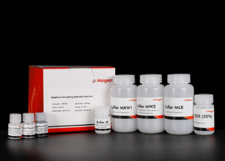
Kit Contents
Contents
1292750
12927200
Purification Times
50
200
MagPure Particles G
20 ml
80 ml
MagBind Particles (selection particles)
14 ml
58 ml
Selection Solution
100 ml
400 ml
Proteinase K
300 mg
1.2 g
Protease Dissolve Buffer
25 ml
100 ml
Buffer SDS(20%)
15 ml
60 ml
Buffer MLK
300 ml
3 x 450 ml
Buffer BST1
225 ml
2x 450 ml
Buffer MKW1
225 ml
2x 450 ml
Buffer MW2*
50 ml
2x 100 ml
Buffer AE
10 ml
30 ml
Storage and Stability
MagPure Particles G, MagBind Particles and Proteinase K should bestored at 2–8°C upon arrival. However, short-term storage (up to 12 weeks) at room temperature (15–25°C) does not affect their performance. The remaining kit components can be stored dry at roomtemperature (15–25°C) and are stable for at least 18 months underthese conditions.The entire kit can be stored at 2–8°C, but in this case buffers should be redissolved before use. Make sure that all buffers are at room temperature when used.
Other Products
Bis-propargyl-PEG2
Product Info
Document
Product Info
Bis-propargyl-PEG2 is a homobifunctional PEG linker with two propargyl groups. The propargyl groups forms triazole linkage with azide-bearing compounds or biomolecules via copper catalyzed Click Chemistry. Reagent grade, for research purpose. Please contact us for GMP-grade inquiries.
Document
Bis-propargyl-PEG2 is a homobifunctional PEG linker with two propargyl groups. The propargyl groups forms triazole linkage with azide-bearing compounds or biomolecules via copper catalyzed Click Chemistry. Reagent grade, for research purpose. Please contact us for GMP-grade inquiries.
Apoptosis is an essentially normal physiological process that removes now redundant, cells, particularly during embryonic development and early growth. In adult animals the process removes cells that are irreparable. The apoptotic process is also involved in many major diseases such as cancer, where transformed tumour cells have their apoptotic process disabled, permitting cell cycling to continue unchecked. In contrast some forms of senile dementia may result from excessive apoptotic induction of neural cells.
The apoptotic process in mammalian cells is a rapid event (2‐4 hours). Within this short time span an apparently viable cell can be quietly dismantled, to disappear leaving no visible trace of its former existence.
How is apoptosis detected or measured?
An apoptosis cascade of activators, effectors and regulators has been identified. This in turn led to a range of apoptosis assays being devised to detect and monitor these events. Some laboratories will employ two distinct assays, one selected to detect early (initiation) apoptotic events, while a second assay will target a later (execution) event. Apoptosis assays, based on methodology, can be classified into four major inter‐linked groups:
[1] DNA fragmentation (electrophoresis and nick end labelling, TUNEL).
[2] Apoptotic proteases (fluorescently labelled antibodies to the caspases).
[3] Flow cytometric analysis (FACS, incorporating other group assays).
Biocolor’s APOPercentage assay is based on the latter. Further information can be found under the ‘Mode of Action’ Tab.
How does APOPercentage detect apoptosis?
The mammalian cell membrane has been described as a semi‐fluid mosaic structure, composed of phospholipids with a diverse group of inserted proteins and some cholesterol. The phospholipids are the major components of the membrane and are arranged in the form of a ‘bi‐layer’; which is asymmetric in composition, structure, and function.
To ensure normal transmembrane functions the phospholipids must be maintained in an asymmetric composition. The process is regulated by ‘flippases’, which catalyse the active transport of aminophospholipids from the outer to inner monolayer. However, in cells undergoing apoptosis, flippase is overwhelmed by the action of another enzyme, termed ‘floppase’ or ‘scramblase’. The net effect is a scrambling of the phospholipid distribution between the inner and outer monolayers.
Cell membrane changes during apoptosis
The APOPercentage assay utilises an intense, pink-coloured dye reagent which is taken up during in-vitro culture by apoptosis-committed cells. This uptake occurs at the stage of Phosphatidylserine transmembrane movement, as produced by the flipflop mechanism. Dye uptake continues until blebbing occurs. No further dye can then enter the now defunct cell and the dye that has accumulated within the cell is not released (unlike necrotic cells which release dye).
Since the dye reagent is excluded or not retained by healthy or necrotic cells it therefore acts as a specific label for apoptotic cells.
How are APOPercentage-labelled cells quantified?
Labelled apoptosis cells may then by conveniently analysed by the following methods:
Direct Analysis The intense pink colour of the labelled cells can be visually assessed using brightfield microscopy. Apoptosis in substrate-adherent cell populations is therefore readily quantified using image analysis techniques. This technique is the most sensitive with the ability of detecting one single apoptotic cell per well.
Colorimetry protocol Dye that accumulates within apoptotic cells is released into solution via addition of Dye Release Reagent. The concentration of this intracellular dye is then measured at 550nm using a microplate colorimeter/spectrophotometer.
NB: The APOPercentage assay kit does NOT require the use of a Flow Cytometer.
Limit of Detection
A single cell (via image analysis method)
Detection Method
Colorimetric (550nm) (Endpoint) or Image Analysis based
Measurements per kit
Sufficient for 4×24 well plates or 6×96 well plates
Suitable Samples
Adherent mammalian cells (in-vitro)
APOPercentage kit contents:
1. APOPercentage Dye (1x5ml)
2. Dye Release Reagent (1x150ml)
3. Phosphate Buffered Saline (PBS) (1x120ml)
4. 24-well starter plate.
5. Assay kit manual.
The Colorimetric Protocol requires a Microplate Colorimeter / Spectrophotometer.
Additional 96-well plates will be required for use when reading dye absorbance values.
The Direct Detection Protocol Requires an inverted stage microscope with an attached digital camera.
NB: Additional reagents (typically culture medium and suitable apoptosis treatments) may be required for sample preparation prior to assay. Consult manual or contact us for further details.
Document
The APOPercentage™ Apoptosis kit is a dye-based, colorimetric assay for detection and measurement of apoptosis (programmed cell death) during in-vitro cell culture.