


K-PHYTASE
SKU: 700004328
100 assays per kit
| Content: | 100 assays per kit |
| Shipping Temperature: | Ambient |
| Storage Temperature: | Short term stability: 2-8oC, Long term stability: See individual component labels |
| Stability: | > 2 years under recommended storage conditions |
| Analyte: | Phytase |
| Assay Format: | Spectrophotometer |
| Detection Method: | Absorbance |
| Wavelength (nm): | 360 |
| Signal Response: | Increase |
| Linear Range: | 0.1 to 10 μg of phosphate per assay |
| Limit of Detection: | 1.5 U/L |
| Reproducibility (%): | < 7% |
| Reaction Time (min): | ~ 30 min |
| Application examples: | Animal feeds, phytase activity in cereal, fungal and bacterial phytases. |
| Method recognition: | Novel method |
The Phytase Assay Kit is a simple, quantitative method which can be used to measure phytase activity. Analysis is based on the hydrolysis of phytic acid by phytase and quantitative measurement of the phosphate released. Results are measured using a standard UV-VIS spectrophotometer and do not require the creation of a standard curve. The Phytase method can be used to measure phytase activity in cereal, fungal and bacterial phytases, and can also be used for the analysis of phytase in animal feed samples.
View our complete list of assay kits for enzyme activities.

Advantages

DBCO-NHCO-PEG13-acid is an analog of DBCO-Acid with a hydrophilic PEG spacer arm, which improves water solubility. This reagent is a non-activated building block with enhanced solubility in aqueous media used to derivatize amine-containing molecule through a stable amide bond. DBCO is commonly used for copper-free Click Chemistry reactions. Reagent grade, for research purpose. Please contact us for GMP-grade inquiries.

DBCO-NHCO-PEG13-acid is an analog of DBCO-Acid with a hydrophilic PEG spacer arm, which improves water solubility. This reagent is a non-activated building block with enhanced solubility in aqueous media used to derivatize amine-containing molecule through a stable amide bond. DBCO is commonly used for copper-free Click Chemistry reactions. Reagent grade, for research purpose. Please contact us for GMP-grade inquiries.

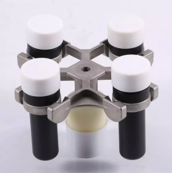
Ideal magnet for all of your low volume needs. From Low elution 96-well PCR applications to 384-well, the X396 has you covered. No need to purchase two separate plates anymore. 96-well volumes as low as 5 µL are achieved from 96-well, PCR plates on the inside of the magnet, while the outside of each magnet will pull beads to well sides in 384-well plates.
ANSI/ SBS Footprint (127.75mm x 85.50mm) to fit into any automated liquid handling robot on bottom. ANSI/SBS footprint on top to accept all common microplates.
Integrated Cushion base for maximum recovery. Helps aid in set-up, robot positioning inconsistencies, and labware consumable inconsistencies
Features include solid aluminum alloy construction and hard coat anodized finish for years of trouble-free use, and compatible with any magnetic beads
A040523
384-Well PCR
Maximum – 39 µL
Minimum – 5 µL
__________
96-Well PCR
Maximum – 60 µL (0.2 mL for some beads)
Minimum – 4 µL
Ideal magnet for all of your low volume needs. From Low elution 96-well PCR applications to 384-well, the X396 has you covered. No need to purchase two separate plates anymore. 96-well volumes as low as 5 µL are achieved from 96-well, PCR plates on the inside of the magnet, while the outside of each magnet will pull beads to well sides in 384-well plates.