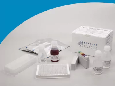
Name of Product Acanthocheilonema viteae – IgG ELISA Catalog Number AF 9400 Short Info This ELISA kit is for the quantitative detection of IgG antibodies against various filiarial nematodes in human serum
This product is manufactured by Bordier Affinity Products in Switzerland and distributed in Germany exclusively by Milenia Biotec.
Method/Platform ELISA in microplate format Range/Assay Sensivity pNPP, λ=405 nm Test Principle Specific antibodies in the sample bind to Acanthocheilonema viteae antigens sensitized on microtiter plates. The presence of parasite specific antibodies is detected with a Protein A alkaline phosphatase conjugate.
Detail
Name of Product
Acanthocheilonema viteae – IgG ELISA
Catalog Number
AF 9400
Short Info
This ELISA kit is for the quantitative detection of IgG antibodies against various filiarial nematodes in human serum
This product is manufactured by Bordier Affinity Products in Switzerland and distributed in Germany exclusively by Milenia Biotec.
Method/Platform
ELISA in microplate format
Range/Assay Sensivity
pNPP, λ=405 nm
Test Principle
Specific antibodies in the sample bind to Acanthocheilonema viteae antigens sensitized on microtiter plates. The presence of parasite specific antibodies is detected with a Protein A alkaline phosphatase conjugate.
Okadaic Acid (OA) is a one of the diarrhetic shellfish poisons (DSP) produced by dinoflagellate genera Dinophysis and Prorocentrum. There are several chemically different toxins associated with DSP.
They are lipophilic and polyether compounds and can be divided into three main groups:
Acidic toxins
Neutral toxins
Other toxins 2 Contamination of shellfish with OA has been associated with harmful algae blooms throughout the world.
In humans, DSP causes dose-dependent symptoms of diarrhea, nausea, and vomiting. The action levels established by the FDA for OA is 200ppb. The EU has established a level of 160 ppb of OA or its equivalent.
The Attogene Okadaic acid ELISA kit enables international and government regulatory agencies, food manufacturers and processors, as well as quality assurance organizations to detect OA in food, feed, fish, and environmental samples of concern.
Okadaic acid is the causative agent of Diarrhetic Shellfish Poisoning (DSP).
FDA and EPA Safety Levels in Regulations and Guidance – 0.16 mg/kg for Clams, mussels, oysters, and whole and roe-on scallops, fresh, frozen, or canned. – National Shellfish Sanitation Program Guide for the Control of Molluscan Shellfish.
Document
Competitive ELISA for the quantitative analysis of Okadaic Acid (DSP) Format: 96-well microtiter plate (12 test strips of 8 wells) Okadaic acid is a potent neurotoxin and phosphatase inhibitor from dinoflagellate black sponges that are associated with seafood poisonings.
Extraction RNA Nucleic Acid Reagent With 12 Months Shelf Life
Product Info
Document
Product Info
Product Description
Nucleic Acid Extraction Reagent – RNA Extraction Kit with 12 Months Shelf Life
Product Description:
Amp-future Bio’s Nucleic Acid Extraction Reagent is a RNA extraction kit that provides the highest quality nucleic acid extraction. The RNA extraction kit is an automated nucleic acid extractor which allows for quick and easy RNA isolation. The reagent is in liquid form and has a shelf life of 12 months, making it a reliable and durable product. With the Nucleic Acid Extraction Reagent, Amp-future Bio provides the accuracy and reliability of a professionally designed RNA extraction kit.
Features:
Product Name: RNA Nucleic Acid Extraction Reagent
Brand: Amp-future Bio
Form: Liquid
Shelf Life: 12 Months
Application: RNA Extract
Keywords: RNA Extraction Kit
Technical Parameters:
Property
Value
Form
Liquid
Product Name
RNA Nucleic Acid Extraction Reagent
Application
RNA Extraction
Brand
Amp-future Bio
Shelf Life
12 Months
Composition
5ml*4 tubes
Applications:
Amp-future Bio’s Nucleic Acid Reagent is an advanced automated nucleic acid extraction solution designed to efficiently extract RNA from biological samples while maintaining quality and accuracy. It is made in China and has a 12-month shelf life. It comes in a liquid form and is suitable for use in automated nucleic acid extractors, allowing for automatic and highly efficient extraction of nucleic acid from various sample types. The reagent is also designed to be easy to use, making it an ideal solution for laboratories that require fast and reliable results. This reagent is a great choice for any research or clinical laboratory that needs efficient and accurate nucleic acid extraction.
Support and Services:
Technical Support and Services for Nucleic Acid Reagent We provide technical support and services for Nucleic Acid Reagents. Our experts are available to help you identify the right reagents for your application, answer any questions, and provide assistance on the use and optimization of these reagents. Our experts are also available to provide advice on troubleshooting and protocol optimization. We offer a range of services to make ordering and stocking of reagents easy and convenient.
We provide:
Expert advice on the selection of the right reagents for your application
Free samples for evaluation
Bulk and custom orders
Technical support and troubleshooting assistance
Secure and reliable online ordering
Fast and convenient shipping options
Please contact us for more information about our technical support and services for nucleic acid reagents.
Document
Amp-future Bio’s Nucleic Acid Extraction Reagent is a RNA extraction kit that provides the highest quality nucleic acid extraction. The RNA extraction kit is an automated nucleic acid extractor which allows for quick and easy RNA isolation. The reagent is in liquid form and has a shelf life of 12 months, making it a reliable and durable product. With the Nucleic Acid Extraction Reagent, Amp-future Bio provides the accuracy and reliability of a professionally designed RNA extraction kit.