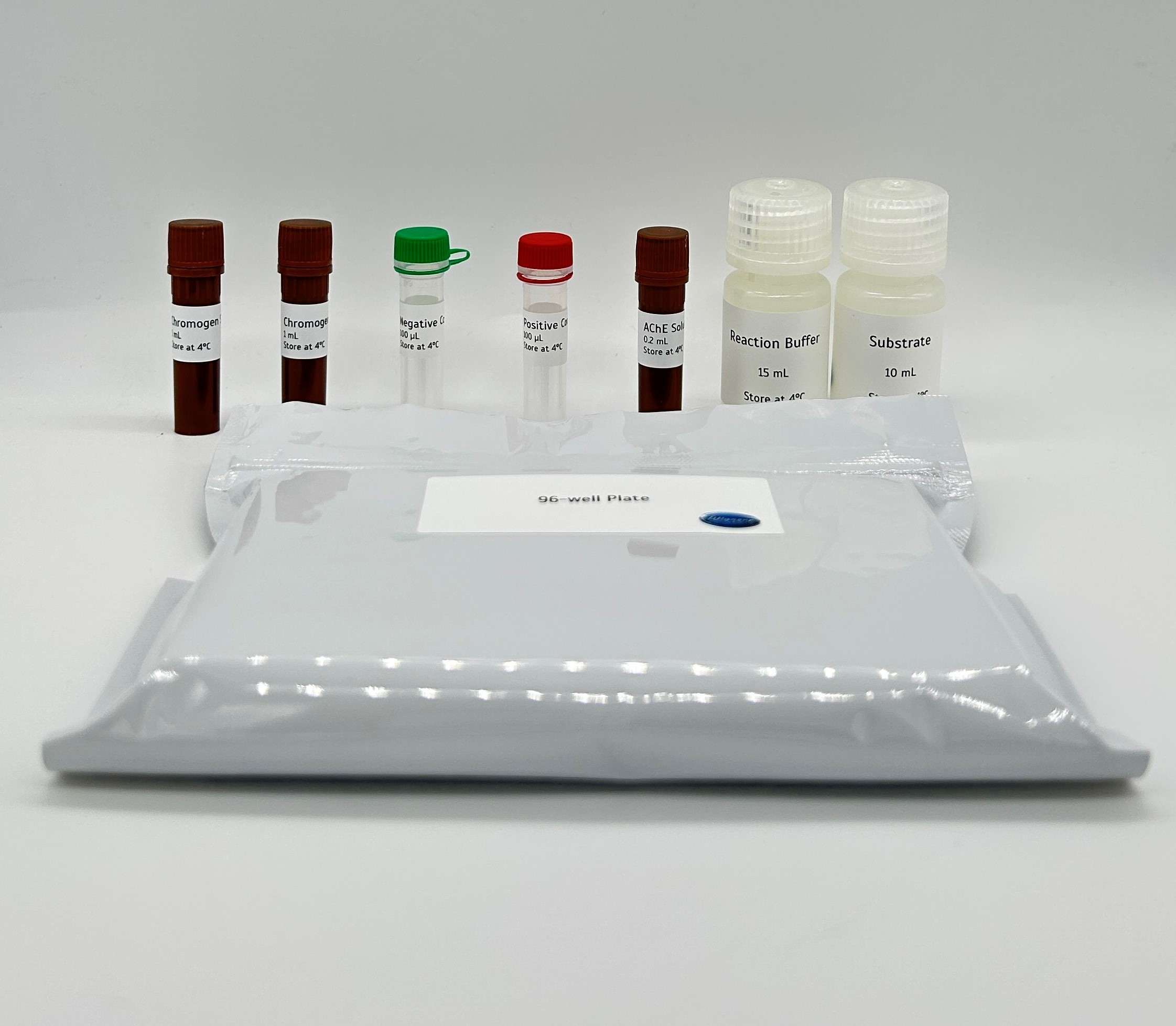
The Acetylcholinesterase Assay Kit provides a convenient method for the detecting AChE activity and screening for inhibitors. The kit uses DTNB to quantify the thiolcholine produced from the hydrolysis of acetylthiolcholine by AChE.
Detail
The Acetylcholinesterase Assay Kit provides a convenient method for the detecting AChE activity and screening for inhibitors.
The kit uses DTNB to quantify the thiolcholine produced from the hydrolysis of acetylthiolcholine by AChE.
AchE Inhibitor Screening Kit (Colorimetric) provides a rapid, simple, sensitive, and reliable test suitable for high-throughput screening of AchE inhibitors.
ACETYLCHOLINESTERASE (EC 3.1.1.7, AChE), also known as RBC cholinesterase, is found primarily in the blood and neural synapses. AChE catalyzes the hydrolysis of the neurotransmitter acetylcholine into choline and acetic acid, a reaction necessary to allow a cholinergic neuron to return to its resting state after activation. Inhibition of the enzyme leads to acetylcholine accumulation, hyperstimulation of nicotinic and muscarinic receptors, and disrupted neurotransmission. AChE inhibition is an important target for the management of Alzheimer’s disease. In addition to Alzheimer’s disease, AChE inhibitors have been useful in the diagnosis or treatment of diseases such as glaucoma, myasthenia gravis, bladder distention, and more.
The Attogene Acetylcholinesterase Inhibitor Assay is based on an improved Ellman method, in which thiocholine produced by the action of acetylcholinesterase forms a yellow color with 5,5′-dithiobis(2-nitrobenzoic acid). The intensity of the product color, measured at 412nm, is proportionate to the enzyme activity in the sample.
Other Products
Biotin-PEG4-C1-alkyne
Product Info
Document
Product Info
Biotin-PEG4-C1-alkyne is azide reactive biotinylation reagent, PEG4 arm increase aqueous solubility of the reagent. Reagent grade, for research purpose. Please contact us for GMP-grade inquiries.
Document
Biotin-PEG4-C1-alkyne is azide reactive biotinylation reagent, PEG4 arm increase aqueous solubility of the reagent. Reagent grade, for research purpose. Please contact us for GMP-grade inquiries.
Cat.# 20106S, 20106L: Size range 450-750 bp (ideal for NGS library size selection)
Product Info
Document
Product Info
The series of DNA Size Selection Kits (Magnetic Beads) were developed for DNA size selection using magnetic beads. A total of 11 kits are available, with different selection ranges spanning from 50 bp to over 10 kb. The kits provide a simple and quick approach for the enrichment of a specific range of DNA fragments. The kit workflow allows double-sided or single-sided size selection for specific size cutoffs.
Gel images of different ranges of size selection. Sheared human genomic DNA was used as input.
.
DNA size selection is a selective capture of DNA fragments of a specific range of size for next-generation sequencing (NGS) library preparations, PCR, ChIP assay, DNA ligations, endonuclease digestions, adapter removal, and other genomics and molecular biology applications. DNA size selection is preferred after NGS library prep in most of the cases. The NGS library preparation is related to the quality of the sequencing data. Precise NGS library size selection can increase sequencing efficiency, improve data quality, and reduce costs.
There are two types of sequencing technologies: short-read sequencing and long-read sequencing. Short-read sequencing uses DNA libraries that contain small insert DNA fragments of similar sizes, usually several hundred base pairs. The sequencing efficiency can be improved if the DNA size selection is in the right range. Cat.# 20104S and 20104L are the best kits for NGS library size selection of illumina paired-end 100 (PE100) sequencing with 100-200 bp library inserts; Cat.# 20105S and 20105L are the best kits for NGS library size selection of illumina paired-end 150 (PE150) sequencing with 150-300 bp library inserts; and Cat.# 20106S and 20106L are the best kits for NGS library size selection of illumina paired-end 300 (PE300) sequencing with 300-600 bp library inserts.
Long-read sequencing uses a large DNA fragment as input and makes very long reads. Usually, library size selection is preferred to remove smaller fragments. Cat.# 20110S and 20110L are the best kits for long-read sequencing size selection with DNA sizes >5 kb, and Cat.# 20111S and 20111L are the best kits for long-read sequencing size selection with DNA sizes >10 kb.
The magnetic beads technology uses paramagnetic particles, also known as SPRI (Solid Phase Reversible Immobilization) beads, to bind DNA reversibly and selectively. DNA fragments can be size-selected and purified by changing the properties of the magnetic beads or SPRI beads. The magnetic beads can easily separate the beads-binding DNA from the contaminants and unwanted components in the samples. The samples after DNA size selection are free of contaminants such as buffer components, enzymes, proteins, salts, dNTPs, primers, and adapters. Our proprietary magnetic beads reagents improve yield, selectivity, and reproducibility.
Specific DNA fragments at a certain length range can be purified simply using magnetic separation with different beads components, avoiding tedious and time-consuming gel extraction and column-based purification. The magnetic beads method is popular for common DNA size selection, including library size selection. The first beads-binding step, referred to as the right-side clean-up, removes large DNA fragments. The large DNA fragments are bound to the beads and are discarded. The desired DNA fragments in the supernatant are transferred to a new well, and new beads are added to the supernatant for the second beads-binding, referred to as the left-side clean-up. The double-size selected DNA fragments are eluted after ethanol rinsing.
DNA size selection with dual clean-ups.
.
A single clean-up is needed for DNA size selection with large fragments. In this case, only the large DNA fragments are bound to the beads. The selected larger DNA fragments are eluted after ethanol rinsing.
DNA size selection with single clean-up for >5 kb and >10 kb DNA.
.
Features of DNA size selection and library size selection
High specificity and high recovery of size selection
11 selection ranges are available, including 5 ranges for NGS library size selection
50-100 bp
100-200 bp
200-500 bp
250-350 bp: ideal for illumina PE100 sequencing
300-450 bp: ideal for illumina PE150 sequencing
450-750 bp: ideal for illumina PE300 sequencing
500-1000 bp
1-3 kb
1-5 kb
>5 kb: ideal for long-read sequencing
>10 kb: ideal for long-read sequencing
Fast and simple
20-min protocol
No gel purification required
No columns required
No centrifugation required
Efficient removal of contaminants and unwanted components
Mucin 6 (MUC6) is a glycoprotein expressed in mucous neck cells, pyloric glands of the antrum, epigastric and bronchial epithelium, and in Müller ducts of the endocervix and urethral epithelium. Anti-MUC6 is useful for differentiating fetal, precancerous, and cancerous colonic mucosa from normal colon, as the antibody does not stain the latter. Anti-MUC6 stains the gastric epithelial surface of normal human gastrointestinal tract.