Basic Nucleic Acid DNA Amplification Kit 48 Reactions 20mins
Facebook
X
Pinterest
Email
Detail
Product Description
Unlocking The Potential Of Nucleic Acid Amplification With DNA Amplification Kit (Basic)
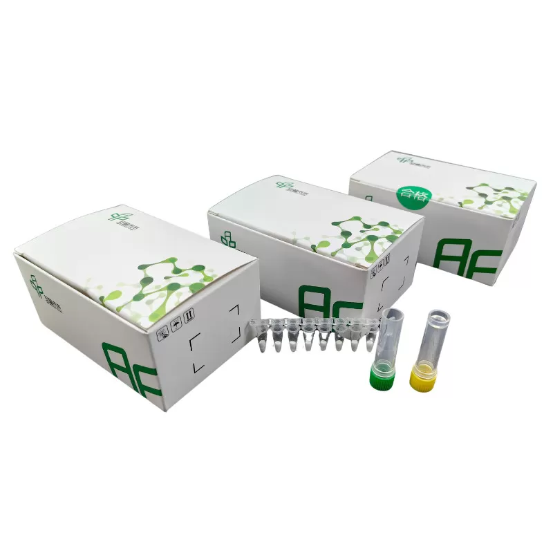
Product Detail
Kit Storage and term of Validity
Storage term: stored at ≤-20℃,keep away from light, avoid heavy weight and repeated freezing and thawing.
Term of Validity: 14 months
Isothermal nucleic acid Principle Summary
The kit is based on rapid nucleic acid amplification technology at room temperature and constant temperature, its principle is that at room and constant temperature, the recombinase and primer form a protein/single-stranded nucleotide complex Rec/ssDNA, and invade the double-stranded DNA template with the help of auxiliary proteins and single-stranded binding protein SSB; then form a D-loop region at the invasion point and start to scan the DNA duplex, after finding the target region complementary to the primer and disintegration of the complex Rec/ssDNA, the polymerase also binds to the 3′ end of the primer to start the chain extension.
Isothermal nucleic acid Product Features
1/ High sensitivity and specificity, short reaction time.
2/ The reagent form is freeze-dried, stable and easy to operate.
3/ The reaction can be operated by metal bath and water bath pot without purchasing expensive PCR apparatus.
Technical Parameters:
Parameters
Details
Product Name
DNA Isothermal Amplification Kit Basic
Manufacturer
Amp-future
Storage Temperature
-20°C
Kit Components
Enzymes, Buffers ,Reagents
Packaging
48 Tests/box
Detection Limit
500-1000copies/µL
Shipping
ICE
Test Time
5-20mins
Isothermal nucleic acid Applications
Suitable for DNA isothermal rapid amplification kit(Basic type)
Primer: Require pair of nucleotide primers with the length of 25-35 bp.
DNA basic kit reaction temperature is 39 to 42℃ and time is 5-20 minutes.
Notes
1/ Please avoid nucleic acid contamination and set blank control during reaction due to the high sensitivity of the kit.
2/ Please take out the required quantity of MIRA reaction units for the experiment, and put the rest under storage conditions when performing the experiment.
Other Products
Cell Culture Plate 96 Wells
Product Info
Document
Product Info
Cell Culture Plate 96 Wells
Cell Culture Plate provide excellent smoothness and uniformity based on accurate molding technology. By such property, clear view can be available when examine with a microscope.
Specifications: 6wells,
12wells, 24wells., 48wells, 96wells
PRODUCT FEATURES
The product is made of medical grade USP CLASS VI polymer polystyrene
The product is made under a 100,00- class dust-free manufacturing site
Two kinds of product line up are providing.
For adherent cell culture: Initial adherence and proliferative property of cells via hydrophilic surface treatment.
For suspension cell culture: The surface is resistant to cell adherence, which minimizes damage or loss of cell.
Gamma radiation sterilization
Non-Pyrogenic, DNase/Rnase free.
Document
Cell Culture Plate provide excellent smoothness and uniformity based on accurate molding technology. By such property, clear view can be available when examine with a microscope.
3,4-Dibromo-Mal-PEG4-amide-DBCO is a PEG linker containing a dibromomaleimide group and a terminal DBCO group. The dibromomaleimide group allows for two points of attachement because both of the bromine atoms can be substituted. The DBCO groups is commonly used for copper-free Click Chemistry reactions due to its strain promoted high energy. PEG linkers are hydrophilic moieties, therefore the attachment of a PEG linker to a compound increases it’s water solubility properties in aqueous media. Reagent grade, for research purpose.
Document
3,4-Dibromo-Mal-PEG4-amide-DBCO is a PEG linker containing a dibromomaleimide group and a terminal DBCO group. The dibromomaleimide group allows for two points of attachement because both of the bromine atoms can be substituted. The DBCO groups is commonly used for copper-free Click Chemistry reactions due to its strain promoted high energy. PEG linkers are hydrophilic moieties, therefore the attachment of a PEG linker to a compound increases it’s water solubility properties in aqueous media. Reagent grade, for research purpose.
Apoptosis is an essentially normal physiological process that removes now redundant, cells, particularly during embryonic development and early growth. In adult animals the process removes cells that are irreparable. The apoptotic process is also involved in many major diseases such as cancer, where transformed tumour cells have their apoptotic process disabled, permitting cell cycling to continue unchecked. In contrast some forms of senile dementia may result from excessive apoptotic induction of neural cells.
The apoptotic process in mammalian cells is a rapid event (2‐4 hours). Within this short time span an apparently viable cell can be quietly dismantled, to disappear leaving no visible trace of its former existence.
How is apoptosis detected or measured?
An apoptosis cascade of activators, effectors and regulators has been identified. This in turn led to a range of apoptosis assays being devised to detect and monitor these events. Some laboratories will employ two distinct assays, one selected to detect early (initiation) apoptotic events, while a second assay will target a later (execution) event. Apoptosis assays, based on methodology, can be classified into four major inter‐linked groups:
[1] DNA fragmentation (electrophoresis and nick end labelling, TUNEL).
[2] Apoptotic proteases (fluorescently labelled antibodies to the caspases).
[3] Flow cytometric analysis (FACS, incorporating other group assays).
Biocolor’s APOPercentage assay is based on the latter. Further information can be found under the ‘Mode of Action’ Tab.
How does APOPercentage detect apoptosis?
The mammalian cell membrane has been described as a semi‐fluid mosaic structure, composed of phospholipids with a diverse group of inserted proteins and some cholesterol. The phospholipids are the major components of the membrane and are arranged in the form of a ‘bi‐layer’; which is asymmetric in composition, structure, and function.
To ensure normal transmembrane functions the phospholipids must be maintained in an asymmetric composition. The process is regulated by ‘flippases’, which catalyse the active transport of aminophospholipids from the outer to inner monolayer. However, in cells undergoing apoptosis, flippase is overwhelmed by the action of another enzyme, termed ‘floppase’ or ‘scramblase’. The net effect is a scrambling of the phospholipid distribution between the inner and outer monolayers.
Cell membrane changes during apoptosis
The APOPercentage assay utilises an intense, pink-coloured dye reagent which is taken up during in-vitro culture by apoptosis-committed cells. This uptake occurs at the stage of Phosphatidylserine transmembrane movement, as produced by the flipflop mechanism. Dye uptake continues until blebbing occurs. No further dye can then enter the now defunct cell and the dye that has accumulated within the cell is not released (unlike necrotic cells which release dye).
Since the dye reagent is excluded or not retained by healthy or necrotic cells it therefore acts as a specific label for apoptotic cells.
How are APOPercentage-labelled cells quantified?
Labelled apoptosis cells may then by conveniently analysed by the following methods:
Direct Analysis The intense pink colour of the labelled cells can be visually assessed using brightfield microscopy. Apoptosis in substrate-adherent cell populations is therefore readily quantified using image analysis techniques. This technique is the most sensitive with the ability of detecting one single apoptotic cell per well.
Colorimetry protocol Dye that accumulates within apoptotic cells is released into solution via addition of Dye Release Reagent. The concentration of this intracellular dye is then measured at 550nm using a microplate colorimeter/spectrophotometer.
NB: The APOPercentage assay kit does NOT require the use of a Flow Cytometer.
Limit of Detection
A single cell (via image analysis method)
Detection Method
Colorimetric (550nm) (Endpoint) or Image Analysis based
Measurements per kit
Sufficient for 4×24 well plates or 6×96 well plates
Suitable Samples
Adherent mammalian cells (in-vitro)
APOPercentage kit contents:
1. APOPercentage Dye (1x5ml)
2. Dye Release Reagent (1x150ml)
3. Phosphate Buffered Saline (PBS) (1x120ml)
4. 24-well starter plate.
5. Assay kit manual.
The Colorimetric Protocol requires a Microplate Colorimeter / Spectrophotometer.
Additional 96-well plates will be required for use when reading dye absorbance values.
The Direct Detection Protocol Requires an inverted stage microscope with an attached digital camera.
NB: Additional reagents (typically culture medium and suitable apoptosis treatments) may be required for sample preparation prior to assay. Consult manual or contact us for further details.
Document
The APOPercentage™ Apoptosis kit is a dye-based, colorimetric assay for detection and measurement of apoptosis (programmed cell death) during in-vitro cell culture.