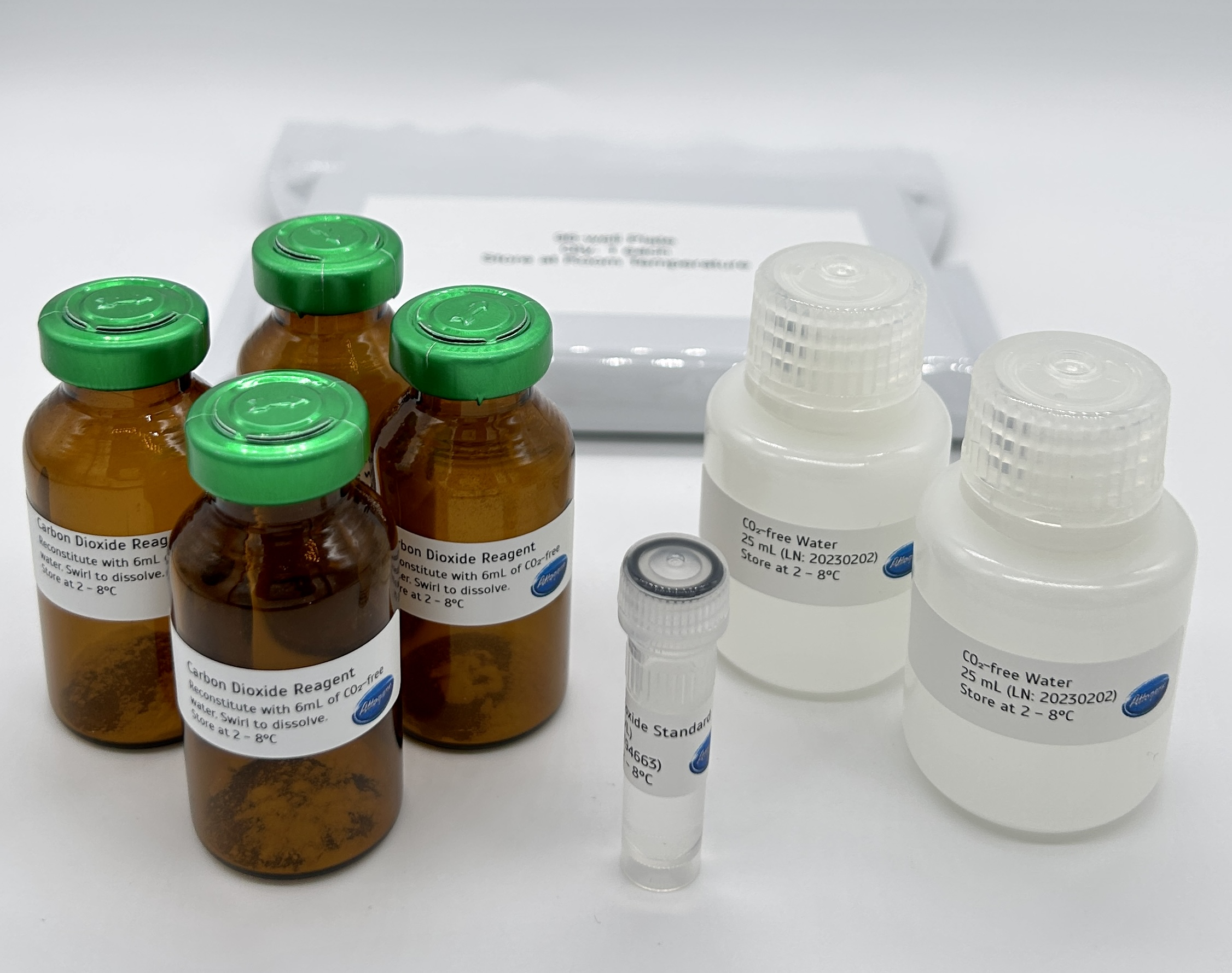
Attogene’s Carbon Dioxide Enzymatic Assay Kit is a simple, direct method for measuring Carbon Dioxide levels in the environment. The assay uses a coupled enzyme assay to detect CO2 (as HCO3-) as follows. In the first step, the bicarbonate condenses with phosphoenol pyruvate to form oxalate (and phosphoric acid); this reaction is catalyzed by the enzyme Phosphoenolpyruvate Decarboxylase, PEPC. The oxalate is then enzymatically reduced by the enzyme Malate Dehydrogenase (using an NADH cofactor) to form malate and NAD+.
Other Products
R6672C MagPure Pathogen DNA/RNA Enrich Kit (tNGS)
Product Info
Document
Product Info
Introduction
This kit is suitable for extracting total pathogen nucleic acid from a variety of clinical samples such as blood, serum, plasma, swab soaking solution, fluid accumulation and homogenate solution. This kit is designed to remove host cells background nucleic acid and enrich pathogen nucleic acid (including viral/bacterial/fungal DNA/RNA) from the sample. Purified DNA/RNA is ready for downstream applications such as PCR, virus detection, tNGS and other related experiments.
Details
Specifications
Features
Specifications
Main Functions
Extract Pathogen RNA/DNA from 0.5-1.5ml whole blood, plasma, serum, body fluid, homogenate suspension, culture solution, cell suspension, soaking solution or concentrate pathogen solution for tNGS application, remove host background nucleic acid.
Applications
Real Time PCR, biochip analysis, NGS
Products
Pathogen DNA / RNA
Purification method
Polydisperse magnetic beads
Purification technology
Magnetic beads technology
Process method
Manual or automatic
Sample type
whole blood, plasma, serum, body fluid, homogenate suspension, culture solution, cell suspension, soaking solution or concentrate pathogen solution
Sample amount
0.5 – 1.5 ml
Adaptive instrument
Nucleic acid extractor, pipetting workstation
Principle
This product is based on the purification method of high binding magnetic particles. The sample is lysed and digested under the action of lysate and Protease. After adding magnetic particles and binding solution, DNA/RNA will be adsorbed on the surface of magnetic particles, and impurities such as proteins will be removed without adsorption. The adsorbed particles were washed with washing solution to remove proteins and impurities, washed with ethanol to remove salts, and finally DNA/RNA was eluted by Buffer NFW.
Kit Contents
Contents
R667200C
R667202C
Purification Times
24 Preps
96 Preps
2ml Bead Tube (0.4g)
24
96
DNase I (Powder)
10 mg
15 mg
DNase Buffer
5 ml
20 ml
Protease Dissolve Buffer
3 ml
8 ml
Lysis Buffer LBX1
40 ml
180 ml
Buffer TL
5 ml
20 ml
Proteinase K
24 mg
120 mg
MagBind Particles N9
1.2 ml
5 ml
Buffer MLB
30 ml
120 ml
Buffer MW1*
13 ml
110 ml
Buffer MW2*
10 ml
50 ml
Buffer AVE
10 ml
20 ml
Storage and Stability
Proteinase K, DNase I powder and MagPure Particles N9 should be stored at 2–8°C upon arrival. However, short-term storage (up to 8 weeks) at room temperature (15–25°C) does not affect their performance. The remaining kit components can be stored at room temperature (15–25°C) and are stable for at least 18 months under these conditions.
Document
This kit is suitable for extracting total pathogen nucleic acid from a variety of clinical samples such as blood, serum, plasma, swab soaking solution, fluid accumulation and homogenate solution. This kit is designed to remove host cells background nucleic acid and enrich pathogen nucleic acid (including viral/bacterial/fungal DNA/RNA) from the sample. Purified DNA/RNA is ready for downstream applications such as PCR, virus detection, tNGS and other related experiments.
A highly efficient, easily automated PCR purification system that delivers superior quality DNA with no salt carryover. Requiring no centrifugation or filtration. This kit can be easily used in manual and automated 96 or 384-well formats.
Details
Specifications
Features
Specifications
Main Functions
Recover 100-400μl DNA/ RNA from PCR products / enzymatic reaction solution / or crude DNA / RNA
Applications
Automatic sequencing, enzyme digestion, PCR and labeling
The MagPure method contains magnetic particles in an optimized binding buffer to selectively bind DNA fragments 100bp and larger toparamagnetic beads. Excess primers, nucleotides, salts, and enzymes can be removed using a simple washing procedure.
Advantages
High recovery efficiency – up to 80% DNA recovery
Kit Contents
Contents
D500301
D500302
D500303
Purification Times
50 Preps
500 Preps
5000 Preps
Buffer AL
10 ml
60 ml
550 ml
Buffer BD*
5 ml
25 ml
2 x 100 ml
MagPure RNA Particles
1.2 ml
12 ml
120 ml
RNase Free Water
5 ml
30 ml
250 ml
Storage and Stability
MagPure RNA Particles should be stored at 2-8°C upon arrival. However, short-term storage (up to 12 weeks) at room temperature (15-25°C) does not affect its performance. The remaining kit components can be stored dry at room temperature (15-25°C) and are stable for at least 18 months under these conditions. The entire kit can be stored at 2-8°C, but in this case buffers should be redissolved before use. Make sure that all buffers are at room temperature when used.
Document
A highly efficient, easily automated PCR purification system that delivers superior quality DNA with no salt carryover. Requiring no centrifugation or filtration. This kit can be easily used in manual and automated 96 or 384-well formats.