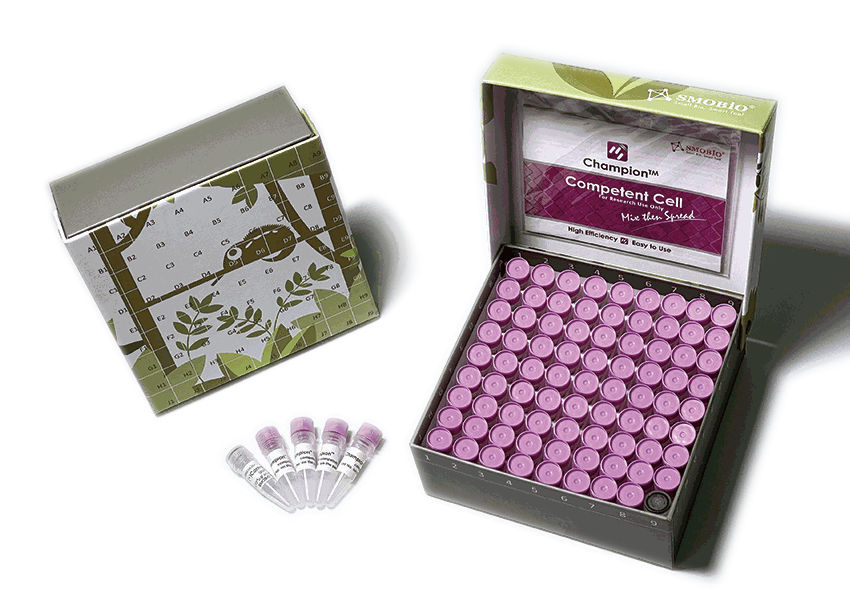
Champion™ Competent Cells are chemically competent cells, which were prepared by SMOBIO to make E. coli perform excellent transformation efficiency. Standard transformation protocol is recommended for large plasmids or non-ampicillin selection. Time-saving transformation protocol is recommended for simple and rapid transformation. Champion™ Competent Cells are one of the fastest and simplest ready-to-use competent cell products in the world.
Detail
General information
Champion™ Competent Cells are chemically competent cells, which were prepared by SMOBIO to make E. coli perform excellent transformation efficiency. Standard transformation protocol is recommended for large plasmids or non-ampicillin selection. Time-saving transformation protocol is recommended for simple and rapid transformation. Champion™ Competent Cells are one of the fastest and simplest ready-to-use competent cell products in the world.
Kit contents
Champion™ Competent Cells
pUC19 Control Plasmid (5 μl, 10-4 μg/μl)
Champion™ Transformation Protocol Card
Shipping condition
Throughout the shipping process, the temperature is maintained under -70°C.
Storage and expiration
Champion™ Competent Cells must be stored between -70°C to -80°C. Subsequent freeze-thaw cycles will reduce transformation efficiency. If high efficiency is required for the experiment, do not use aliquots that have gone through several freeze-thaw cycles. The efficiency of Champion™ Competent Cells lasts for 1 year with proper storage.
For rapid, sensitive and accurate screening of potential Tyrosinase inhibitors
Tyrosinase (EC 1.14.18.1) is a copper-binding enzyme that is expressed across a vast range of species ranging from bacteria and fungi to mammals. It is involved in two sequential reactions of the melanin synthesis pathway: first being the hydroxylation of a monophenol and second the conversion of an ortho-diphenol to a quinone. Quinone then undergoes a series of reactions including polymerization to form melanin.
Tyrosinase is of great interest to the agriculture industry since it causes browning of fruits, vegetable, and mushrooms, as well as to the cosmetic industry as it causes skin darkening. Development and screening of tyrosinase inhibitors, therefore, is very useful for conditions such as hyperpigmentation and melasma. Tyrosinase activity is significantly increased in melanoma. Therefore, the detection of tyrosinase activity could be promising as a specific diagnostic test for melanoma and may be useful in monitoring patient response to melanoma treatments.
This Tyrosinase Activity Assay Kit is a simple one-step, plate-based assay for the measurement of tyrosinase activity in biological samples. In this assay, tyrosinase catalyzes the conversion of a phenolic substrate to a Quinone intermediate, which reacts with the tyrosine enhancer forming a highly stable chromophore with absorbance at 520 nm. The assay can detect as low as 30 μU Tyrosinase in biological samples.
Document
Highly reproducibility Highly Sensitive Assay to Screen for Tyrosinase Activity Stable formulation of ready to use Reaction Facilitator (tyrosinase)
The 16S V1-V2 Library Preparation Kit for Illumina consists of the reagents and components required for library preparation of the 16S V1-V2 amplicon libraries to be used for next-generation sequencing on Illumina platforms. All molecular reagents including primers, enzyme mixes, indexes, and buffers are provided. Instructions for PCR clean up with the AMPure XP Magnetic Beads (supplied by customer) are also included for rapid purification of nucleic acid products generated at two steps of the workflow. The library prep workflow could be used for purified DNA inputs from different sources including stool, soil, water, saliva, plant, urine, skin swab, vaginal swab, cheek swab, nasal swab, plasma/serum, tongue swab, gum swab, and others.
The 16S V1-V2 Library Preparation Kit for Illumina has a streamlined procedure that reduces the handling time such that the library prep procedure can be completed in approximately 4 hours (see diagram below). Input DNA is first subjected to targeted PCR to amplify the V1-V2 region of the DNA encoding 16S rRNA. The post-PCR reaction is then cleaned up using AMPure XP beads. Dual index primers are then added using a limited-cycle PCR. The indexed amplicons flanked by 5′ and 3′ barcoded adaptors are then cleaned using AMPure XP beads. The libraries are then ready for quantification, pooling and sequencing.
Storage Conditions and Product Stability Norgen’s 16S V1-V2 Library Prep Kit for Illumina is shipped as one kit box (for the 24 prep kit) or two sub-component kits (for the 96 prep kit). All kits should be stored at -20°C upon arrival.
All kit components should remain stable for at least 1 year when stored at the specified storage conditions.
African Swine Fever Virus (ASFV) is a widespread disease which infects members of the pig family(Suidae). Anumberoftick species are believed to be the vector for the disease,as well as being transmitted by raw pork and pig excrement [1]. After firstly being identified in Kenya in 1921, ASFV became endemic in sub-Saharan Africa, with regular outbreaks being reported across Europe, Asia and South America throughout the century [2]. More recently the virus was introduced in Georgia and spread throughout the region, as well as mass outbreaks occurring in China in 2018 [3]. ASFVistheonlymemberoftheAsfaridaefamily.ItisalargeenvelopeddoublestrandedDNA virus of icosahedral morphology with an average diameter of 200nm and isolates contain genomes between 170-190Kbp encoding for up to 167 open reading frames [2]. The morphology of ASFV consist of several concentric domains. An inner core contains the nucleoid coated with a thick protein layered core shell, which is surrounded by an inner lipid envelope , all of which is encompassed by the capsid [2]. ASFV begins its replication cycle in the nucleus of infected cells before moving to the cytoplasm where the majority of the replication takes place [2]. Gene transcription is highly regulated, with distinct classes of mRNA identified to accumulate at early, intermediate and late transcripts of the virus [2]. The disease induces acute haemorrhagic disease within its hosts, causing high fevers and skin haemorrhages, with death often occurring within ten days of clinical symptoms appearing [4].
References: 1: The Centre for Food Security and Public Health (2015), African Swine Fever. 2: Galindo, I. and Alonso, C., 2017. African swine fever virus: a review. Viruses, 9(5), p.103. 3: Zhou, X., Li, N., Luo, Y., Liu, Y., Miao, F., Chen, T., Zhang, S., Cao, P., Li, X., Tian, K. and Qiu, H.J., 2018. Emergence of African swine fever in China, 2018. Transboundary and emerging diseases, 65(6), pp.1482-1484. 4: Gallardo, C., Ademun, A.R., Nieto, R., Nantima, N., Arias, M., Martín, E., Pelayo, V. and Bishop, R.P., 2011. Genotyping of African swine fever virus (ASFV) isolates associated with disease outbreaks in Uganda in 2007. African Journal of biotechnology, 10(17), pp.3488-3497.
Document
Exceptional value for money Rapid detection of all clinically relevant subtypes Positive copy number standard curve for quantification Highly specific detection profile High priming efficiency Broad dynamic detection range (>6 logs) Sensitive to < 100 copies of target
Accurate controls to confirm findings