[CD1021] SMOChem™ Deoxynucleotide (dNTP) Mix, 25 mM each (100 mM total), 500 µl x 6
Facebook
X
Pinterest
Email
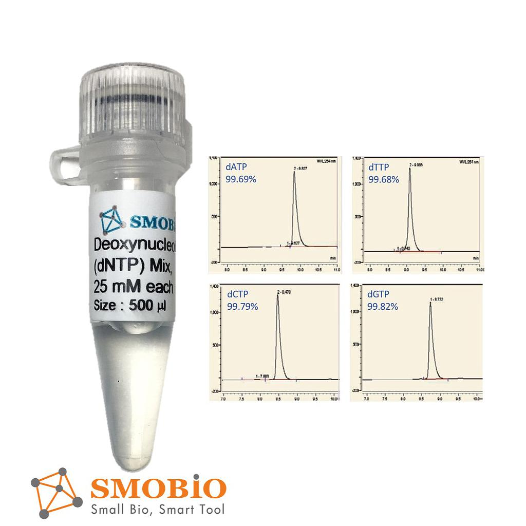
The SMOChem™ Deoxynucleotide (dNTP) Mix is an aqueous solution that contains an equimolar solution of ultrapure dATP, dCTP, dGTP and dTTP, each at a concentration of 25 mM at pH 8.5. The dNTP Mix is designed for many different molecular biology applications that involved in DNA synthesis or labeling, such as PCR, real-time PCR, DNA sequencing, reverse transcription, primer extension, and etc. The dNTP Mix is free of exo-deoxyribonuclease and endo-deoxyribonuclease as well as ribonuclease activity. The dNTP Mix offers the possibility to reduce the number of pipetting steps and the risk of reaction set up errors.
Detail
Description
The SMOChem™ Deoxynucleotide (dNTP) Mix is an aqueous solution that contains an equimolar solution of ultrapure dATP, dCTP, dGTP and dTTP, each at a concentration of 25 mM at pH 8.5. The dNTP Mix is designed for many different molecular biology applications that involved in DNA synthesis or labeling, such as PCR, real-time PCR, DNA sequencing, reverse transcription, primer extension, and etc. The dNTP Mix is free of exo-deoxyribonuclease and endo-deoxyribonuclease as well as ribonuclease activity. The dNTP Mix offers the possibility to reduce the number of pipetting steps and the risk of reaction set up errors.
Short term stability: 2-8oC, Long term stability: See individual component labels
Stability:
> 2 years under recommended storage conditions
Analyte:
Formaldehyde
Assay Format:
Spectrophotometer, Microplate, Auto-analyser
Detection Method:
Absorbance
Wavelength (nm):
340
Signal Response:
Increase
Linear Range:
0.1 to 14 μg of formaldehyde per assay
Limit of Detection:
0.033 mg of formaldehyde per test or 0.016 mg/mL of formaldehyde in a sample treated as per the standard procedure
Limit of Quantification:
0.11 mg of formaldehyde per test or 0.054 mg/mL of formaldehyde in a sample treated as per the standard procedure
Reproducibility (%):
~ 3%
Reaction Time (min):
~ 15 min
Application examples:
Environmental samples, food and beverage samples.
The Formaldehyde Assay Kit provides a simple robust method for the measurement of formaldehyde.
Note for Content: The number of manual tests per kit can be doubled if all volumes are halved. This can be readily accommodated using the MegaQuantTM Wave Spectrophotometer (D-MQWAVE).
Features of the sDNASEQ E.coli Residual DNA Quantitation kit include:
Simpler and Rapid
Only three steps will be need for Sample Preparation and All components of the Sample Preparation Kit can be stored at room temperature.
Only one Reagent for qPCR;
Only 1.5 hours will be needed for the whole test.
Accurate
Perfect amplification curve, good amplification efficiency and good precision.
Highly sensitive quantitation using proven TaqMan™ real-time qPCR technology.
Limit of Detection (LoD): 1 fg/μL; Limit of Quantification (LoQ):5 fg/μL.
The recovery rate of different concentration samples in the linear range is between 70% and 130%
Kit Performance
Fig 1. Only three steps will be need for Sample Preparation and only 20 minitutes will be taken for Sample Preparation.
Fig 2. Seven concentration samples of 5fg/μL, 10fg/μL, 20fg/μL, 30fg/μL, 300fg/μL, 3pg/μL, 30pg/μL, 300pg/μL were detected. CV of each concentration was < 30%, Regression coefficient associated with standard solutions was 0.99975, and amplification efficiency was 100.068%.
Fig 3. Four concentration samples of 5fg/μL, 10fg/μL, 20fg/μL, and 30fg/μL were detected, and 10 multiple holes were detected for each concentration. The detection values of 5fg/μL and above were CV <30%.
Fig 4. DNA recovery can be determined by including samples spiked with known DNA amounts which are prepared from the corresponding DNA standards. Typically, the range for this value varies from 70% to 130%.
Fig 5. Only one Reagent for qPCR MIX.
Document
Note: Price not include shipment & duty, contact us to get full quote. The resDNASEQ E.coli Residual DNA Quantitation kit is designed for the quantification of residual DNA from E. coli, in cell lines which are used for production of biopharmaceutical products. The resDNASEQ E.coli Residual DNA Quantitation kit use TaqManTM quantitative PCR to perform rapid, specifc quantitation of femtogram levels of residual host-cell or plasmid DNA. The kit was developed to meet the sensitivity requirements defined by WHO (10 ng E. coli DNA per therapeutic dose).
Magen’s HiPure columns are prepared by high quality glass fiber filter membrane as raw materials through membrane cutting, membrane release, ring release, ring pressing, gland, weighing and other processes. HiPure nucleic acid adsorption columns have the characteristics of long-term stability and high binding capacity. Experiments show that the highest binding capacity and binding efficiency of HiPure nucleic acid adsorption columns are basically unchanged when stored at room temperature for 4 years.
The series of nucleic acid columns produced by Magen Biotech are based on carefully selected imported glass fiber membranes (GF/B, GF/D, GF/F). Columns production processes such as polypropylene injection molding materials, injection molding process, and downstream membrane packing and compression rings are strictly controlled. This is to ensure that the column has extremely high adsorption capacity and long-term stability. Compared with conventional products on the market, Magen’s columns are with varieties, and binding rate will not change when stored at room temperature for 4 years.
Details
Specifications
Features
Specifications
Recommended application
Plasmid Medium Yield preparation
Preservation conditions
Room temperature
Stability
Up to 4 years
Filter membrane
High quality glass fiber filter GF/B, 8 layers
Membrane aperture
1.0 μm
Maximum binding yield of plasmid
250 μg
Maximum yield of alcohol mediated Binding
1 mg
Plasmid Yields
Up to 0.25mg
Single liquid carrying capacity of column
4 ml
Minimum elution volume
500 μl
Withstand centrifugal force
5,000 x g
Centrifuge
Lowspeed centrifuge for 15ml centrifuge tubes, >3000 x g, swing-out Rotor, or Fixed Angle Rotor
Adsorption Mechanism
Based on the negatively charged DNA skeleton, it has a high affinity for positively charged glass fibers. In high salt and ethanol solutions, DNA/RNA binds to glass fiber and interacts with hydrophilic matrix on silica through hydrogen bond. DNA/RNA is tightly bound. All pollutants can be removed by washing solution. At high salt concentration, nucleic acids selectively bind to silica gel membrane, while other pollutants, mainly proteins, are removed by membrane washing.
Ordering information
CAT.No.
Product Name
Package
C13121
HiPure DNA Midi Column III (8 x GF/B)with 15ml Collection Tubes
100/Bag
Purchase Guide
Item No.
Product Name
Membrane type/number of layers
Collection tubes
Plasmid DNA binding capacity (Physical adsorption)
Note: GF/B pore size is for 1.0μM glass fiber membrane; GF/F pore size is for 0.7μm glass fiber membrane.
Document
Magen’s HiPure columns are prepared by high quality glass fiber filter membrane as raw materials through membrane cutting, membrane release, ring release, ring pressing, gland, weighing and other processes. HiPure nucleic acid adsorption columns have the characteristics of long-term stability and high binding capacity. Experiments show that the highest binding capacity and binding efficiency of HiPure nucleic acid adsorption columns are basically unchanged when stored at room temperature for 4 years.