
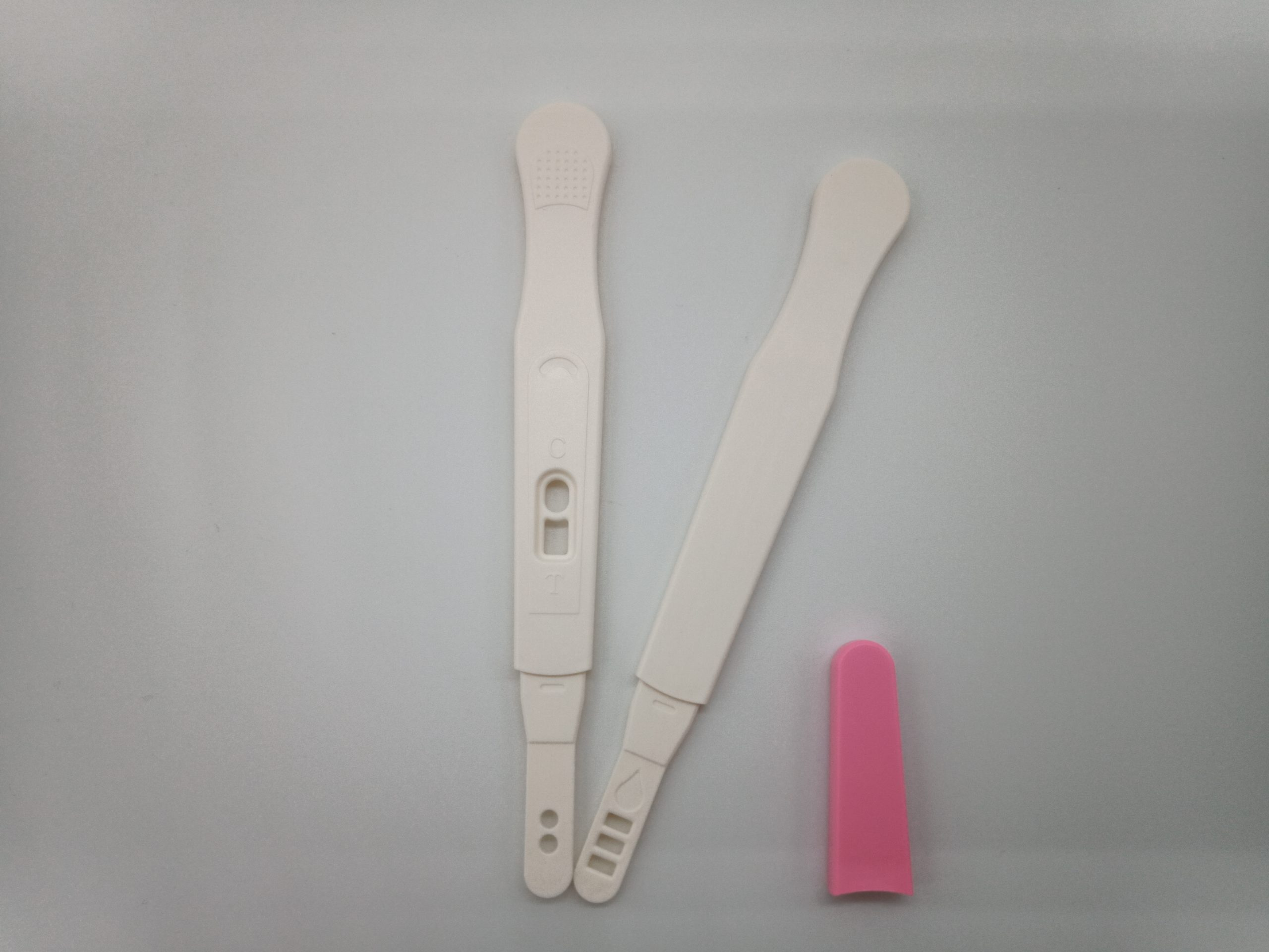
Description
- Appearance: White/pink
- • Length of strip: 86mm
- • Width of strip: 4.5mm
- • Length of device: 136mm
- • Width of device: 16mm


Blood samples contain rich DNA, including mitochondrial DNA, genomic DNA, circulating DNA (mostly released into blood after tumor cell apoptosis) in white blood cells, as well as parasitic viral or microbial DNA. These DNA are important parameters in clinical testing or diagnosis, which are also valuable materials for medical research. There are three main issues with extracting DNA from blood samples:
1. The sample is highly infectious, posing great harm to operators and the environment.
2. The source of DNA is complex and aportion of the nucleic acid, such as viral DNA or free DNA, may be lost during the operation, leading to downstream detection failure;
3. Blood sample contains a large amount of impurities and inhibitory factors.
Currently there are many methods available for extracting DNA from whole blood samples, such as phenol chloroform extraction, salting out method, etc. However, these methods require pre-treatment of blood sample, which removes red blood cells and isolate white blood cells in the first step. Due to the requirement that it cannot inactivate or kill pathogens during the process of removing red blood cells, the waste liquid (red blood cell lysate) and consumables may be contaminated by pathogens and become infectious, posing a danger to the entire laboratory environment and operators. In addition, during the process of removing red blood cells, useful nucleic acid information such as viruses, microorganisms, or circulating DNA is also lost, leading to experiment or detection failures.
The HiPure Blood DNA Kits series provided by Magen Company uses silica gel column purification technology, which can directly lyse whole blood samples without the need for white blood cell separation. Whole blood samples are directly mixed with lysates and proteases, resulting in the inactivation of pathogens, greatly reducing the infectivity, environmental pollution, and the chance of operators being infected. Due to the direct lysis and digestion of samples, except lymphocyte DNA, other circulating DNA as well as DNA from viruses and microorganisms, can also be recovered.
This product provides fast and easy methods for purification of total DNA for reliable PCR and Southern blotting. Total DNA (e.g., genomic, viral, mitochondrial) can be purified from tissue, whole blood, plasma, serum, buffy coat, bone marrow, other body fluids, lymphocytes, cultured cells.
Specifications
| Features | Specifications |
| Main Functions | Isolation total DNA from blood, tissue, culture cells, swab, blood spots using 96 plate |
| Applications | PCR, southern bolt and virus detection, etc |
| Purification method | 96 well plate |
| Purification technology | Silica technology |
| Process method | Manual (centrifugation or vacuum) |
| Sample type | Blood, serum, plasma, milk, saliva, and other liquid samples and cultured cells |
| Sample amount | |
| Elution volume | |
| Time per run | |
| Liquid carrying volume per column | |
| Binding yield of column |
This product is based on silica column purification. The sample is lysed and digested with lysate and protease, DNA is released into the lysate. Transfer to an adsorption column. Nucleic acid is adsorbed on the membrane, while protein is not adsorbed and is removed with filtration. After washing proteins and other impurities, Nucleic acid was finally eluted with low-salt buffer (10mm Tris, pH9.0, 0.5mm EDTA).
| Contents | D311701 | D311702 |
| Purification Times | 1 x 96 | 4 x 96 |
| HiPure gDNA Plate | 1 | 4 |
| 96 well Plate (2.2ml) | 1 | 4 |
| 1.6ml Collection Plate | 1 | 4 |
| 0.5ml Collection Plate | 1 | 4 |
| Silicon Seal Tape | 1 | 4 |
| Seal Film | 5 | 25 |
| Buffer ATL | 30 ml | 100 ml |
| Buffer AL | 30 ml | 100 ml |
| Buffer DW1 | 60 ml | 250 ml |
| Buffer GW2 | 50 ml | 2 x 100 ml |
| Proteinase K | 50 ml | 200 ml |
| Protease Dissolve Buffer | 5 ml | 15 ml |
| Buffer AE | 30 ml | 120 ml |
Storage and Stability
Proteinase K should be stored at 2-8°C upon arrival. However, short-term storage (up to 12 weeks) at room temperature (15-25°C) does not affect their performance. The remaining kit components can be stored at room temperature (15-25°C) and are stable for at least 18 months under these conditions.
| Name | CAT NO | Sample amount | Leukocyte protocol* | Colum type | Elutio volume | Average yield | Time per run |
| HiPure Blood DNA Mini Kit | D3111 | 10-200μl | 1ml | 2ml column | ≥20μl | 5-9μg/200μl | ≤30 minutes |
| HiPure Tissue&Blood DNA Midi Kit | D3113 | 0.2-2ml | 10ml | 1.5ml column | ≥300μl | 20-40μg/1m | ≤80 minutes |
| HiPure Tissue&Blood DNA Maxi Kit | D3115 | 3 -10ml | 10ml | 15ml column | ≥700μl | 20-40μg/1m | ≤90 minutes |
| HiPure Tissue&Blood DNA 96 Kit | D3117 | 1-200μl | 1ml | 96 well plate | 3-8μg/200μl |
Blood samples contain rich DNA, including mitochondrial DNA, genomic DNA, circulating DNA (mostly released into blood after tumor cell apoptosis) in white blood cells, as well as parasitic viral or microbial DNA. These DNA are important parameters in clinical testing or diagnosis, which are also valuable materials for medical research. There are three main issues with extracting DNA from blood samples:

Description
The DM3160 FluoroBand™ 1KB (0.25-10 kb) Fluorescent DNA Ladder is a ready-to-use DNA ladder, which is pre-mixed with high sensitivity DNA binding fluorescent dye and loading dye for direct gel loading. The DNA ladder DM3160 is composed of 13 individual DNA fragments: 10k, 8k, 6k, 5k, 4k, 3k, 2.5k, 2k, 1.5k, 1k, 750, 500, and 250 bp derived from a mixture of PCR products and specifically digested plasmid DNA; these bands can be visualized when illuminated with 470 nm blue light or UV light. This product contains two enhanced bands (3 kb and 1 kb) for easier reference. In addition, two tracking dyes, Xylene cyanol FF and Bromophenol blue which mimic the migration of 4,000 bp and 500 bp dsDNA during electrophoresis are also added for real time monitoring. Real time observation of the electrophoresis is also possible if compatible light source is fitted to the electrophoresis tank.
Features
Source
Phenol extracted PCR products and dsDNA digested with specific restriction enzymes, equilibrated in 10 mM Tris-HCl (pH 8.0) and 10 mM EDTA.
Range
250 ~ 10,000 bp
Concentration
50 µg/ 500 µl
Recommended loading volume
5 µl/ well
Storage
Protected from light
Room temperature for 6 months
4°C for 12 months
-20°C for 24 months
The DM3160 FluoroBand™ 1KB (0.25-10 kb) Fluorescent DNA Ladder is a ready-to-use DNA ladder, which is pre-mixed with high sensitivity DNA binding fluorescent dye and loading dye for direct gel loading. The DNA ladder DM3160 is composed of 13 individual DNA fragments: 10k, 8k, 6k, 5k, 4k, 3k, 2.5k, 2k, 1.5k, 1k, 750, 500, and 250 bp derived from a mixture of PCR products and specifically digested plasmid DNA; these bands can be visualized when illuminated with 470 nm blue light or UV light. This product contains two enhanced bands (3 kb and 1 kb) for easier reference. In addition, two tracking dyes, Xylene cyanol FF and Bromophenol blue which mimic the migration of 4,000 bp and 500 bp dsDNA during electrophoresis are also added for real time monitoring. Real time observation of the electrophoresis is also possible if compatible light source is fitted to the electrophoresis tank.

| Clone | IHC010 |
| Source | Mouse Monoclonal |
| Positive Control | Melanoma, Skin Melanocytes |
| Dilution Range | 1:200 |
SRY (Sex Determining Region Y)-Box 10 (SOX-10), also known as transcription factor SOX-10, is a nuclear transcription factor that acts in regulation of embryonic development and in the specification and differentiation of cells of melanocytic lineage. SOX-10 is diffusely expressed in neurofibromas and schwannomas, and mutations in the SOX-10 gene are linked to Waardenburg-Shah and Waardenburg-Hirschsprung disease. Anti-SOX-10 has been shown to be sensitive for conventional, spindled, and desmoplastic melanoma, and has been used to detect metastatic melanoma and nodal capsular nevus in sentinel lymph nodes.