FOR US ONLY Rapid detection and exclusive to the COVID-19 strain Does not detect other related coronavirus strains High priming efficiency Accurate controls to confirm extraction, and assay validity Lyophilised components for ambient shipping Highly specific detection profile This test has been authorized by FDA under an EUA for use by authorized laboratories.
Detail
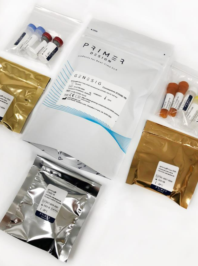
This test has been authorized by FDA under an EUA for use by authorized laboratories.
The Primerdesign Ltd COVID-19 genesig® Real-Time PCR assay is a real-time RT-PCR assay intended for the qualitative detection of nucleic acid from SARS-CoV-2 in oropharyngeal swab specimens from patients suspected of COVID-19 by their healthcare provider. Testing is limited to laboratories certified under the Clinical Laboratory Improvement Amendments of 1988 (CLIA), 42 U.S.C. §263a, to perform high complexity tests.
This product provides high quality purification of total DNA from whole blood, plasma, serum, buffy coat, or other body fluids, lymphocytes and cultured cells. There is no need to use toxic phenol chloroform extraction or time-consuming alcohol precipitation. The extraction process finish in 60 minutes. Purified DNA includes genomic DNA, mitochondrial DNA, viral DNA (e.g. HBV), or DNA from other parasitic microorganisms. The obtained DNA can be directly used in PCR, viral DNA detection and other experiments.
This kit can use on manual protocol or 96 channel automated extraction system.
Details
Specifications
Features
Specifications
Main Functions
Isolation total DNA from 200μl whole blood
Applications
PCR, southern bolt and virus detection, etc
Purification technology
Magnetic beads technology
Process method
Manual or automatic
Sample type
Anticoagulant blood, concentrated blood, buffy coat, lymphocytes and cultured cells
Sample amount
200μl
Elution volume
≥50μl
Time per run
≤60 minutes
Principle
This product is based on the purification method of high binding magnetic particles. The sample is lysed and digested under the action of lysate and Protease. DNA is released into the lysate. After adding magnetic particles and binding solution, DNA will be adsorbed on the surface of magnetic particles, and impurities such as proteins will be removed without adsorption. The adsorbed particles were washed with washing solution to remove proteins and impurities, washed with ethanol to remove salts, and finally DNA was eluted by Elution Buffer.
Advantages
High binding force – suitable for handling DNA rich samples, such as whole blood, buffy coat, concentrated blood, etc.
Fast – polydisperse magnetic beads, fast magnetic response and short extraction time
High purity – the obtained DNA can be directly used for second-generation sequencing, PCR based detection, gene bank, etc.
Automatic – saving time and labor and safer
Kit Contents
Contents
D631101
D631102
D631103
Purification Times
48
96
480
MagPure Particles
1.2 ml
2.5 ml
11 ml
Proteinase K
24 mg
50 mg
220 mg
Protease Dissolve Buffer
1.8 ml
5 ml
15 ml
Buffer AL
15 ml
30 ml
120 ml
Buffer GW1*
22 ml
53 ml
220 ml
Elution Buffer
15 ml
30 ml
100 ml
Storage and Stability
Proteinase K, MagPure Particles should be stored at 2-8°C upon arrival. However, short-term storage (up to 24 weeks) at room temperature (15-25°C) does not affect their performance. The remaining kit components can be stored dry at room temperature (15-25°C) and are stable for at least 18 months under these conditions. The entire kit can be stored at 2-8°C, but in this case buffers should be redissolved before use. Make sure that all buffers are at room temperature when used.
Document
This product provides high quality purification of total DNA from whole blood, plasma, serum, buffy coat, or other body fluids, lymphocytes and cultured cells. There is no need to use toxic phenol chloroform extraction or time-consuming alcohol precipitation. The extraction process finish in 60 minutes. Purified DNA includes genomic DNA, mitochondrial DNA, viral DNA (e.g. HBV), or DNA from other parasitic microorganisms. The obtained DNA can be directly used in PCR, viral DNA detection and other experiments.
This kit can use on manual protocol or 96 channel automated extraction system.
TD4N Table Top 5000rpm Low Speed Laboratory Centrifuge
Product Info
Document
Product Info
TD4N Table Top 5000rpm Low Speed Laboratory Centrifuge
TD4N Features:
1. Microprocessor control, less noisily, it is widely used to qualitative analysis of blood serum, plasma and urea in the fields of hospital, blood center, laboratory and biochemistry.
2. Brushelss motor, free maintenance, no powder pollution, quick in speed up and down.
3. The flexible axle driven system which drive the rotor directly, smooth in operation and small vibration.
4. There are many rotors for your choice, suitable for different specifications meet customers’ different requirements of separation.
5. Micro-computer control system, digital display the RCF, time and speed.
6. Electric lid lock, compact design, super speed and imbalance protection.
TD4N Technical Parameter:
Max. Speed
5000rpm
Speed Accuracy
±20rpm
Max. Volume
6x100ml
Power Supply
AC110V/220V 50HZ/60HZ
Max. RCF
3460xg
Noise
≤55dBA
Timer
0~99min
Net Weight
18Kg
Dimension
540x370x280mm
Certifications
CE, ISO & Calibration report are available.
Warranty
1 Year
Matched Rotors for TD4N:
Order No.
Rotor Type
Max.Speed(rpm)
Max.Volume(ml)
Max.RCF(xg)
4N-1
Swing Rotor
5000
6x10ml
3460
4N-2
Angle Rotor
4000
30×7/5ml
2250
4N-3
Angle Rotor
4000
18×15/10ml
2250
4N-4
Angle Rotor
4000
24x10ml
2200
4N-5
Angle Rotor
4000
12×15/7/5ml
2150
4N-6
Angle Rotor
4000
12x20ml
2200
4N-7
Angle Rotor
5000
6x15ml
2540
4N-8
Angle Rotor
5000
12x15ml
3080
4N-9
Angle Rotor
5000
4x50ml
2520
4N-10
Angle Rotor
5000
6x50ml
2850
4N-11
Angle Rotor
5000
4x100ml
2630
4N-12
Angle Rotor
5000
6x100ml
3130
Document
TD4N is a table top centrifuge widely used to qualitative analysis of blood serum, plasma and urea in the fields of hospital, blood center, laboratory and biochemistry.
This ELISA kit is intended for the quantitative detection of IgG antibodies against Echinococcus granulosus (and Echinococcus multilocularis) in human serum.
This product is manufactured by Bordier Affinity Products in Switzerland and distributed in Germany exclusively by Milenia Biotec.
Method/Platform
ELISA in microplate format
Range/Assay Sensivity
pNPP, λ=405 nm
Test Principle
Specific antibodies in the sample bind to Echinococcus granulosus Antigens sensitized on microtiter plates. The presence of parasite specific antibodies is detected with a Protein A – alkaline phosphatase conjugate.
Document
12 x 8 strips (96 tests)
This ELISA kit is intended for the quantitative detection of IgG antibodies against Echinococcus granulosus (and Echinococcus multilocularis) in human serum.
This product is manufactured by Bordier Affinity Products in Switzerland and distributed in Germany exclusively by Milenia Biotec.