
Introduction
Usage: For cultivating, enumerating and preserving non-fastidious bacteria. (ISO)

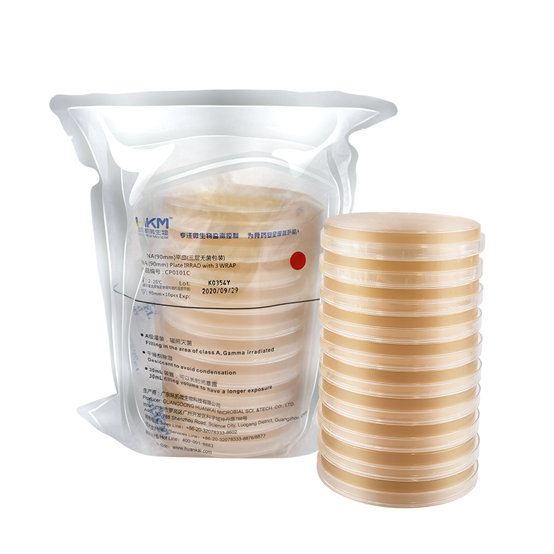
Usage: For cultivating, enumerating and preserving non-fastidious bacteria. (ISO)

As this is a 2 gene kit, we recommend purchase of 2 of the accompanying RT-qPCR master mix reagent: oasig Lyophilised OneStep RT-qPCR Master Mix 150 reactions.
Norovirus is known to cause acute gastroenteritis. It is a small (27-38 nm), round, nonenveloped RNA virus belonging to the Caliciviridae family and is responsible for over 80% of non-bacterial outbreaks of gastroenteritis in the world. It affects individuals of all ages, with a distinct seasonal link to winter. It has a genome of 7.6 kb that is positive sense and has a single stranded linear confirmation. It encodes a major structural protein (VP1) of about 58 to 60 kDa and a minor capsid protein (VP2). Transmission occurs predominantly through ingestion of contaminated water, food and airborne transmission, as well as contact with contaminated surfaces. The ease with which norovirus is transmitted and the low infectious dose required to establish an infection results in extensive outbreaks in numerous environments, such as hospitals, hotels and schools. There is no antiviral drug available to treat this infection and little is known about its pathogenicity. However, it has been observed that the virus can be taken up by enterocytes where translation of viral nonstructural proteins can occur; it damages and alters intestinal microvilli, leaving them blunt and broadened, thus inhibiting absorption; it causes crypt cell hyperplasia and also leads to apoptosis of enterocyctes. An incubation period of 24-48 hours is usual. Infection is characterized by the acute onset of nausea, vomiting, abdominal cramps, aching limbs, raised temperature and diarrhoea that generally last for about 48 hours. However, more severe and prolonged infection may be observed in children and the elderly. There are five recognized norovirus genogroups, of which three (GI, GII, and GIV) are known to affect humans and, since 2002, variants of the GII.4 genotype have been the most common cause of norovirus outbreaks. There have been 31 different genotypes identified within the genogroups, with a wide degree of genetic variability present even within each genotype.
Exceptional value for money
Rapid detection of all clinically relevant subtypes
Positive copy number standard curve for quantification
Highly specific detection profile
High priming efficiency
Broad dynamic detection range (>6 logs)
Sensitive to < 100 copies of target
Accurate controls to confirm findings


Description
The PM1500 ExcelBand™ All Blue Regular Range Protein Marker is a blue protein standard with 10 pre-stained proteins covering a wide range of molecular weights from 10 to 180 kDa in Tris-Glycine buffer (9 to 170 kDa in Bis-Tris (MOPS) buffer and Bis-Tris (MES) buffer). Proteins are covalently coupled with a blue chromophore, and two reference bands (at 25 kDa and 72 kDa, respectively) are enhanced in intensity when separated on SDS-PAGE (Tris-Glycine buffer).
The PM1500 ExcelBand™ All Blue Regular Range Protein Marker is designed for monitoring protein separation during SDS-polyacrylamide gel electrophoresis, verification of Western transfer efficiency on membranes (PVDF, nylon, or nitrocellulose) and for approximating the size of proteins.
Features
Contents
Approximately 0.1~0.5 mg/ml of each protein in the buffer (20 mM Tris-phosphate (pH 7.5 at 25°C), 2% SDS, 0.2 mM DTT, 3.6 M urea, and 15% (v/v) glycerol).
Quality Control
Under suggested conditions, PM1500 ExcelBand™ All Blue Regular Range Protein Marker resolves 10 major bands in SDS-PAGE (Tris-Glycine buffer, MOPS, and MES buffer) and after Western blotting to nitrocellulose membrane.
Storage
4°C for 3 months
-20°C for long term storage
The PM1500 ExcelBand™ All Blue Regular Range Protein Marker is a blue protein standard with 10 pre-stained proteins covering a wide range of molecular weights from 10 to 180 kDa in Tris-Glycine buffer (9 to 170 kDa in Bis-Tris (MOPS) buffer and Bis-Tris (MES) buffer). Proteins are covalently coupled with a blue chromophore, and two reference bands (at 25 kDa and 72 kDa, respectively) are enhanced in intensity when separated on SDS-PAGE (Tris-Glycine buffer).
The PM1500 ExcelBand™ All Blue Regular Range Protein Marker is designed for monitoring protein separation during SDS-polyacrylamide gel electrophoresis, verification of Western transfer efficiency on membranes (PVDF, nylon, or nitrocellulose) and for approximating the size of proteins.