
Introduction
Place of Origin: Guangdong, China
Warranty: 2 years
Customized support: OEM
Brand Name: HKM
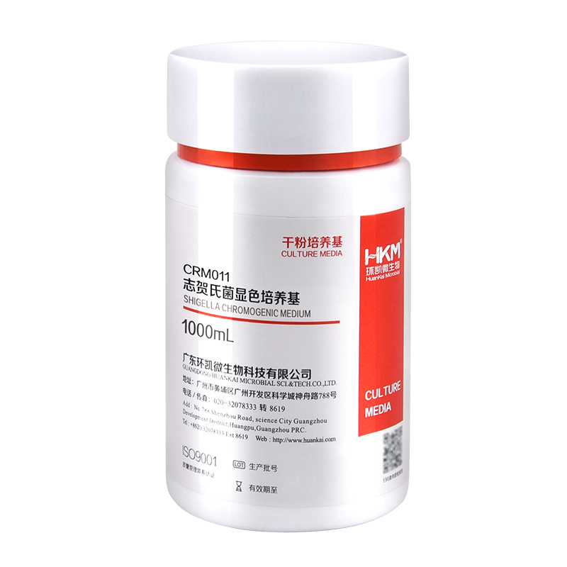
Model Number: CRM011
Reagent grade: Biochemical Reagent
Form: Powder
Color: Light yellow
Type: Shigella Agar

Place of Origin: Guangdong, China
Warranty: 2 years
Customized support: OEM
Brand Name: HKM
Model Number: CRM011
Reagent grade: Biochemical Reagent
Form: Powder
Color: Light yellow
Type: Shigella Agar

This kit provides a fast, reliable and simple procedure for isolating DNA from urine volumes ranging from 3 mL to 25 mL. Both high molecular weight DNA (greater than 1 kb in size; mostly cell associated) and the smaller DNA (150 – 250 bp; derived from the circulation) is effectively isolated and purified with a rapid and convenient spin column protocol. Multiple samples can be processed in 45 minutes. Salts, metabolic wastes, proteins and other contaminants are removed to yield inhibitor-free DNA for use in sensitive applications such as PCR, qPCR, DNA fingerprinting, methylation studies and more. This kit can also be used to isolate DNA from a broad range of viruses.
This kit is fully compatible with Norgen’s Urine Collection and Preservation Tubes.
Norgen’s Urine DNA Isolation Kit Dx (Slurry Format) does not provide a diagnostic result. It is the sole responsibility of the user to use and validate the kit in conjunction with a downstream in vitro diagnostic assay.
NOTE: This product is not available for sale in the United States.
Figure 1 / 3

Click for expanded view
| Kit Specifications – Spin Column | |
| Minimum Urine Input | 3 mL |
| Maximum Urine Input | 25 mL |
| Time to Complete 10 Purifications | < 30 minutes |
| Size of Urine DNA Purified | Large (> 1 kb) and small (150-250 bp) |
Storage Conditions and Product Stability
All solutions should be kept tightly sealed and stored at room temperature. All solutions and plastics can be used until the expiration date specified on their labels. It is recommended to warm Solution A and Solution B for 20 minutes at 60°C if any salt precipitation is observed
| Component | Cat. Dx48800 (50 preps) |
|---|---|
| Solution A | 18 mL |
| Solution B | 30 mL |
| Wash Solution | 22 mL |
| Elution Buffer | 6 mL |
| Mini Filter Spin Columns | 50 |
| Collection Tubes | 50 |
| Elution Tubes (1.7 mL) | 50 |
| Product Insert | 1 |

Apoptosis is an essentially normal physiological process that removes now redundant, cells, particularly during embryonic development and early growth. In adult animals the process removes cells that are irreparable. The apoptotic process is also involved in many major diseases such as cancer, where transformed tumour cells have their apoptotic process disabled, permitting cell cycling to continue unchecked. In contrast some forms of senile dementia may result from excessive apoptotic induction of neural cells.
The apoptotic process in mammalian cells is a rapid event (2‐4 hours). Within this short time span an apparently viable cell can be quietly dismantled, to disappear leaving no visible trace of its former existence.
An apoptosis cascade of activators, effectors and regulators has been identified. This in turn led to a range of apoptosis assays being devised to detect and monitor these events. Some laboratories will employ two distinct assays, one selected to detect early (initiation) apoptotic events, while a second assay will target a later (execution) event. Apoptosis assays, based on methodology, can be classified into four major inter‐linked groups:
[1] DNA fragmentation (electrophoresis and nick end labelling, TUNEL).
[2] Apoptotic proteases (fluorescently labelled antibodies to the caspases).
[3] Flow cytometric analysis (FACS, incorporating other group assays).
[4] Membrane alterations (phosphatidylserine flip).
Biocolor’s APOPercentage assay is based on the latter. Further information can be found under the ‘Mode of Action’ Tab.
The mammalian cell membrane has been described as a semi‐fluid mosaic structure, composed of phospholipids with a diverse group of inserted proteins and some cholesterol. The phospholipids are the major components of the membrane and are arranged in the form of a ‘bi‐layer’; which is asymmetric in composition, structure, and function.
To ensure normal transmembrane functions the phospholipids must be maintained in an asymmetric composition. The process is regulated by ‘flippases’, which catalyse the active transport of aminophospholipids from the outer to inner monolayer. However, in cells undergoing apoptosis, flippase is overwhelmed by the action of another enzyme, termed ‘floppase’ or ‘scramblase’. The net effect is a scrambling of the phospholipid distribution between the inner and outer monolayers.
The APOPercentage assay utilises an intense, pink-coloured dye reagent which is taken up during in-vitro culture by apoptosis-committed cells. This uptake occurs at the stage of Phosphatidylserine transmembrane movement, as produced by the flipflop mechanism. Dye uptake continues until blebbing occurs. No further dye can then enter the now defunct cell and the dye that has accumulated within the cell is not released (unlike necrotic cells which release dye).
Since the dye reagent is excluded or not retained by healthy or necrotic cells it therefore acts as a specific label for apoptotic cells.
Labelled apoptosis cells may then by conveniently analysed by the following methods:
Direct Analysis
The intense pink colour of the labelled cells can be visually assessed using brightfield microscopy. Apoptosis in substrate-adherent cell populations is therefore readily quantified using image analysis techniques. This technique is the most sensitive with the ability of detecting one single apoptotic cell per well.
Colorimetry protocol
Dye that accumulates within apoptotic cells is released into solution via addition of Dye Release Reagent. The concentration of this intracellular dye is then measured at 550nm using a microplate colorimeter/spectrophotometer.
NB: The APOPercentage assay kit does NOT require the use of a Flow Cytometer.
A single cell (via image analysis method)
Colorimetric (550nm) (Endpoint) or Image Analysis based
Sufficient for 4×24 well plates or 6×96 well plates
Adherent mammalian cells (in-vitro)
1. APOPercentage Dye (1x5ml)
2. Dye Release Reagent (1x150ml)
3. Phosphate Buffered Saline (PBS) (1x120ml)
4. 24-well starter plate.
5. Assay kit manual.
The Colorimetric Protocol requires a Microplate Colorimeter / Spectrophotometer.
Additional 96-well plates will be required for use when reading dye absorbance values.
The Direct Detection Protocol Requires an inverted stage microscope with an attached digital camera.
NB: Additional reagents (typically culture medium and suitable apoptosis treatments) may be required for sample preparation prior to assay. Consult manual or contact us for further details.
The APOPercentage™ Apoptosis kit is a dye-based, colorimetric assay for detection and measurement of apoptosis (programmed cell death) during in-vitro cell culture.

N-t-Boc-Aminooxy-PEG4-N-(PEG2-Propargyl) is a click chemistry crosslinker. The propargyl group is reactive with azide-containing compounds or biomolecules through copper catalyzed Click Chemistry to yield a stable triazole linkage. t-Boc-aminooxy can be deprotected under mild acidic conditions. The hydrophilic PEG linker improves solubility in aqueous media.

N-t-Boc-Aminooxy-PEG4-N-(PEG2-Propargyl) is a click chemistry crosslinker. The propargyl group is reactive with azide-containing compounds or biomolecules through copper catalyzed Click Chemistry to yield a stable triazole linkage. t-Boc-aminooxy can be deprotected under mild acidic conditions. The hydrophilic PEG linker improves solubility in aqueous media.