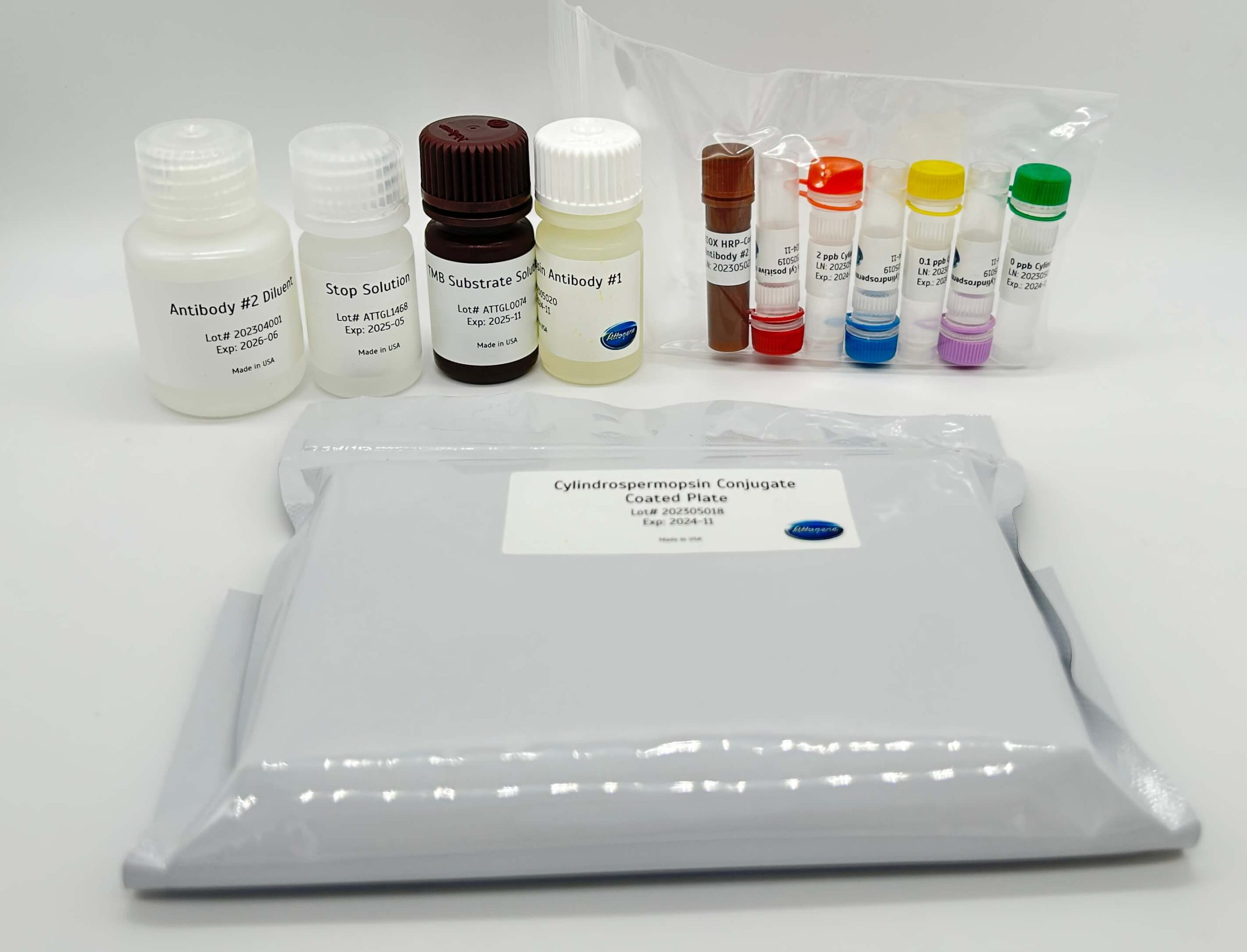
The Cylindrospermopsin plate kit is a competitive enzyme-labeled immunoassay. The Cylindrospermopsin sample extract and calibrators are pipetted into the test wells followed by the Cylindrospermopsin antibody into the test wells to initiate the reaction. During the 30 minutes incubation period, Cylin-drospermopsin from the sample and Cylindrospermopsin antigen compete for binding to the Cylindrosper-mopsin antibody. The Cylindrospermopsin antibody is captured on the walls of the test well. Following this 30-minute incubation, the contents of the wells are removed and the wells are washed to remove any unbound Cylindrospermopsin and free Cylindrospermopsin antibody. After wash, 1X HRP-conjugated Antibody#2 is added for 30 minutes incubation. The wells are washed afterwards, and a clear substrate is then added to the wells and any bound enzyme conjugate causes the conversion to a blue color. Following a 15-minute incubation, the reaction is stopped and the amount of color in each well is read. The color of the unknown samples is compared to the color of the calibrators and the Cylindrospermopsin concentration of the samples is derived.
Format:
• 96-well microtiter plate (12 test strips of 8 wells)
Do not Drink – 0.7 μg/L for bottle fed infants and preschool children, pregnant and nursing woman, elderly immunocompromised and liver conditions.
Do not Drink – 3.0 μg/L for school age children to adults.
Do Not Use – 20 μg/L
EPA Draft Human Health Recreational Ambient Water Quality Criteria to protect human health: 8 μg/L.
Other Products
endo-BCN-PEG3-acid
Product Info
Document
Product Info
endo-BCN-PEG3-acid is a click chemistry linker consisting of a BCN group with a terminal carboxylic acid. The terminal carboxylic acid can react with primary amine groups in the presence of activators (e.g. EDC, or HATU) to yield a amide bond. The BCN group can react with azide-tagged biomolecules. The hydrophilic PEG spacer increases solubility in aqueous media. Reagent grade, for research purpose. Please contact us for GMP-grade inquiries.
Document
endo-BCN-PEG3-acid is a click chemistry linker consisting of a BCN group with a terminal carboxylic acid. The terminal carboxylic acid can react with primary amine groups in the presence of activators (e.g. EDC, or HATU) to yield a amide bond. The BCN group can react with azide-tagged biomolecules. The hydrophilic PEG spacer increases solubility in aqueous media. Reagent grade, for research purpose. Please contact us for GMP-grade inquiries.
Introducing the Fastin Assay Kit: Your Straightforward Solution for Elastin Quantification! Our user-friendly kit utilizes a dye-based method to measure elastin from in-vivo and in-vitro sources. It can be used to quantify various elastin forms, spanning from immature tropoelastin to mature, ‘insoluble’ elastin fibers.
Colorimetric Detection (513nm) (Endpoint)
Understanding Elastin: The Key to Tissue Flexibility
Tissues like lungs and arteries must maintain the ability to stretch and recoil repeatedly throughout an organism’s life. Elastin, a mature protein, is responsible for this elasticity and is usually present as insoluble fibers within the ECM. During development, these fibers are initially formed from a soluble precursor called tropoelastin.
What is the Fastin Assay?
The Biocolor Fastin assay is a user-friendly, dye-based means of quantifying elastins derived from both in-vivo and in-vitro sources. A variety of elastin forms can be assayed, from immature tropoelastin to mature ‘insoluble’ elastin fibres.
Further information on how the assay works can be found on the ‘Mode of Action‘ tab.
A list of suggested sample types can be found under the ‘Assay Specification‘ tab.
How does the Fastin assay detect Elastin?
The Fastin Dye Reagent contains an elastin-binding synthetic porphyrin, TPPS (5,10,15,20-tetraphenyl- 21H,23H-porphine). This affinity of TPPS for elastin was first observed when used as a ‘vital stain’ on live animals. Most tissues took up the dye initially but only elastin retained the TPPS molecules over time. [Winkelman, J. (1962), Cancer Res. 22, 589-596; Winkelman, J & Spicer, S. (1962), Stain Technol. 37, 303-305].
It has been proposed that the elastin binding of TPPS may be due to the retention of the acidic dye (which contains four charged sulfate groups) by the basic amino acid side chain residues of elastin.
How does the Fastin assay work?
Step 1. Incubation of samples containing soluble elastin with the Fastin Dye Reagent causes an elastin-dye complex to form. This insoluble complex then precipiates.
Step 2. Dye-labelled elastin is then isolated by centrifugation and the unbound dye removed. Elastin-bound dye is then eluted and measured spectrophotometrically.
Step 3. The elastin content of unknown samples can be calculated by comparison against a calibration curve prepared using a standard comprising water-soluble elastin (supplied with the kit).
Assay range
50 – 500µg/ml
Limit of Detection
50µg/ml
Detection Method
Colorimetric Detection (513nm) (Endpoint)
Measurements per kit
110 in total (allows a maximum of 48 samples to be run in duplicate alongside a standard curve).
Suitable Samples
In-vivo: tissues and fluids. Insoluble elastin will first require conversion to water soluble α-elastin using the oxalic acid reagents and extraction protocol supplied with the kit.
In-vitro: Elastin produced by cells during 2D/3D cell culture. NB elastin in conditioned cell media is typically below the detection limit of the kit.
Precautions
This kit is designed for research use only. Not for use in diagnostic procedures. Kit requires access to a centrifuge, heated water bath or block, as well as a spectrophotometer or colorimeter capable of absorbance detection at 513nm. Specific sample preparation protocols may require customer to provide further reagents, consult assay manual for further information.
Fastin elastin kit contents:
1. Dye Reagent (1x110ml)
2. α-elastin Reference Standard (1x5ml, 1.0 mg/ml soluble Bovine elastin)
6. 1.5ml micro-centrifuge tubes for dye-labelling reaction.
7. Assay kit manual
NB: Additional reagents may be required for sample preparation prior to assay. Consult manual or contact us for further details.
Document
Introducing the Fastin Assay Kit: Your Straightforward Solution for Elastin Quantification! Our user-friendly kit utilizes a dye-based method to measure elastin from in-vivo and in-vitro sources. It can be used to quantify various elastin forms, spanning from immature tropoelastin to mature, ‘insoluble’ elastin fibers. Colorimetric Detection (513nm) (Endpoint)
MutL Homolog 1 (MLH1) is a protein involved in the mismatch-repair pathway. This protein is commonly associated with hereditary non-polyposis colorectal cancer, as the MLH1 gene is frequently mutated in patients with this cancer. Studies have shown MLH1 to be deficient in a high percentage of patients with microsatellite instability, as well as endometrial and ovarian cancers. Use of Anti-MLH1 is optimized when paired in an IHC panel with MSH6, MSH2, and PMS2. Anti-MLH1 is useful in the detection of MLH1 in a number of normal and neoplastic tissues, and for identifying a loss of MLH1 in tumors that are microsatellite-unstable.