

K-HDBA
SKU: 700004302
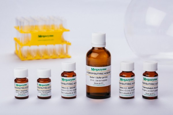
60 assays (manual) / 600 assays (microplate) / 740 assays (auto-analyser)
| Content: | 60 assays (manual) / 600 assays (microplate) / 740 assays (auto-analyser) |
| Shipping Temperature: | Ambient |
| Storage Temperature: | Short term stability: 2-8oC, Long term stability: See individual component labels |
| Stability: | > 2 years under recommended storage conditions |
| Analyte: | D-3-Hydroxybutyric Acid |
| Assay Format: | Spectrophotometer, Microplate, Auto-analyser |
| Detection Method: | Absorbance |
| Wavelength (nm): | 492 |
| Signal Response: | Increase |
| Linear Range: | 0.4 to 12 µg of D-3-hydroxybutyric acid per assay |
| Limit of Detection: | 0.074 mg/L |
| Reaction Time (min): | ~ 6 min |
| Application examples: | Egg, egg products (e.g. egg powder) and other materials (e.g. biological cultures, samples, etc.). |
| Method recognition: | Methods based on this principle have been accepted by EEC |
The D-3-Hydroxybutyric Acid (β-Hydroxybutyrate) Assay Kit is suitable for the specific measurement and analysis of D-hydroxybutyric acid in eggs and egg products and other foods and beverages.
Note for Content: The number of manual tests per kit can be doubled if all volumes are halved. This can be readily accommodated using the MegaQuantTM Wave Spectrophotometer (D-MQWAVE).
Explore more organic acid assay kit products.

Advantages
- Very competitive price (cost per test)
- All reagents stable for > 2 years after preparation
- Very rapid reaction (~ 3 min)
- No wasted diaphorase solution (stable suspension supplied)
- Mega-Calc™ software tool is available from our website for hassle-free raw data processing
- Standard included
- Suitable for manual, microplate and auto-analyser formats


