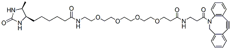
DBCO-PEG4-Desthiobiotin is a PEG linker containing a desthiobiotin group and a DBCO functional group. Desthiobiotin is used for affinity-based applications such as pull-down assays or for ligating with streptavidin proteins while DBCO is a click chemistry handle that quickly reacts with azide groups on target molecules. Desthiobiotin is a sulfur-free analogue of biotin which binds streptavidin with slightly less strength than biotin, which provides it with a soft-release characteristic that is useful for in pull-down assays by minimizing co-elution with endogenous biotinylated molecules. The inclusion of a PEG linker in this molecule improves its aqueous solubility.
Detail
DBCO-PEG4-Desthiobiotin is a PEG linker containing a desthiobiotin group and a DBCO functional group. Desthiobiotin is used for affinity-based applications such as pull-down assays or for ligating with streptavidin proteins while DBCO is a click chemistry handle that quickly reacts with azide groups on target molecules. Desthiobiotin is a sulfur-free analogue of biotin which binds streptavidin with slightly less strength than biotin, which provides it with a soft-release characteristic that is useful for in pull-down assays by minimizing co-elution with endogenous biotinylated molecules. The inclusion of a PEG linker in this molecule improves its aqueous solubility.
Other Products
[TK1000] ExcelTaq™ Klen-Taq DNA Polymerase, 5 U/μl, 500 U
Product Info
Document
Product Info
Description
ExcelTaq™ Klen-Taq DNA Polymerase is a specially blended enzyme mix containing KlenTaq-1 DNA polymerase (a 5’-exo-minus, N-terminal deletion of Taq DNA polymerase) and a small amount of a proofreading DNA polymerase. This unique blending helps to improve the fidelity, yield and processivity of the resultant PCR process. The Klen-Taq is also highly robust, showing high tolerance of varying concentrations of Mg2+; it is highly thermostable and has four times the fidelity compared to Taq DNA polymerase. The ExcelTaq™ Klen-Taq DNA Polymerase is ideal for DNA amplifications 0.5-5 kb in length on genomic DNA, and up to 10 kb on less complex templates.
Features
5’→3′ DNA polymerase activity
3’→5′ exonuclease activity (proofreading)
4× fidelity as compared to Taq DNA polymerase
Thermo-stable: up to 98°C during PCR denaturing step
Robust PCR performance, resistance to variance in PCR conditions
Storage
-20°C for 24 months
Document
ExcelTaq™ Klen-Taq DNA Polymerase is a specially blended enzyme mix containing KlenTaq-1 DNA polymerase (a 5’-exo-minus, N-terminal deletion of Taq DNA polymerase) and a small amount of a proofreading DNA polymerase. This unique blending helps to improve the fidelity, yield and processivity of the resultant PCR process. The Klen-Taq is also highly robust, showing high tolerance of varying concentrations of Mg2+; it is highly thermostable and has four times the fidelity compared to Taq DNA polymerase. The ExcelTaq™ Klen-Taq DNA Polymerase is ideal for DNA amplifications 0.5-5 kb in length on genomic DNA, and up to 10 kb on less complex templates.
N-bis(PEG2-propargyl)-N-(PEG2-amidoPEG1)-N-(bis(PEG2-propargyl) is a branched PEG linker with four terminal alkyne groups. The alkyne groups can react with azides via copper catalyzed Click Chemistry reactions. The PEG units help increase the solubility of the molecule in aqueous environment. Reagent grade, for research purpose. Please contact us for GMP-grade inquiries.
Document
N-bis(PEG2-propargyl)-N-(PEG2-amidoPEG1)-N-(bis(PEG2-propargyl) is a branched PEG linker with four terminal alkyne groups. The alkyne groups can react with azides via copper catalyzed Click Chemistry reactions. The PEG units help increase the solubility of the molecule in aqueous environment. Reagent grade, for research purpose. Please contact us for GMP-grade inquiries.
Aminooxy-PEG1-propargyl is a click chemistry PEG linker containing a propargyl group and an aminooxy group. The aminooxy group is reactive with an aldehyde or ketone group to form a stable oxime linkage. The propargyl group can react with azide-bearing compounds or biomolecules via copper catalyzed azide-alkyne Click Chemistry to yield a stable triazole linkage. Reagent grade, for research purpose. Please contact us for GMP-grade inquiries. Aminooxy compounds are very reactive and sensitive; they cannot be stored for long term. Immediate use (within 1 week) is highly recommended.
Document
Aminooxy-PEG1-propargyl is a click chemistry PEG linker containing a propargyl group and an aminooxy group. The aminooxy group is reactive with an aldehyde or ketone group to form a stable oxime linkage. The propargyl group can react with azide-bearing compounds or biomolecules via copper catalyzed azide-alkyne Click Chemistry to yield a stable triazole linkage. Reagent grade, for research purpose. Please contact us for GMP-grade inquiries. Aminooxy compounds are very reactive and sensitive; they cannot be stored for long term. Immediate use (within 1 week) is highly recommended.