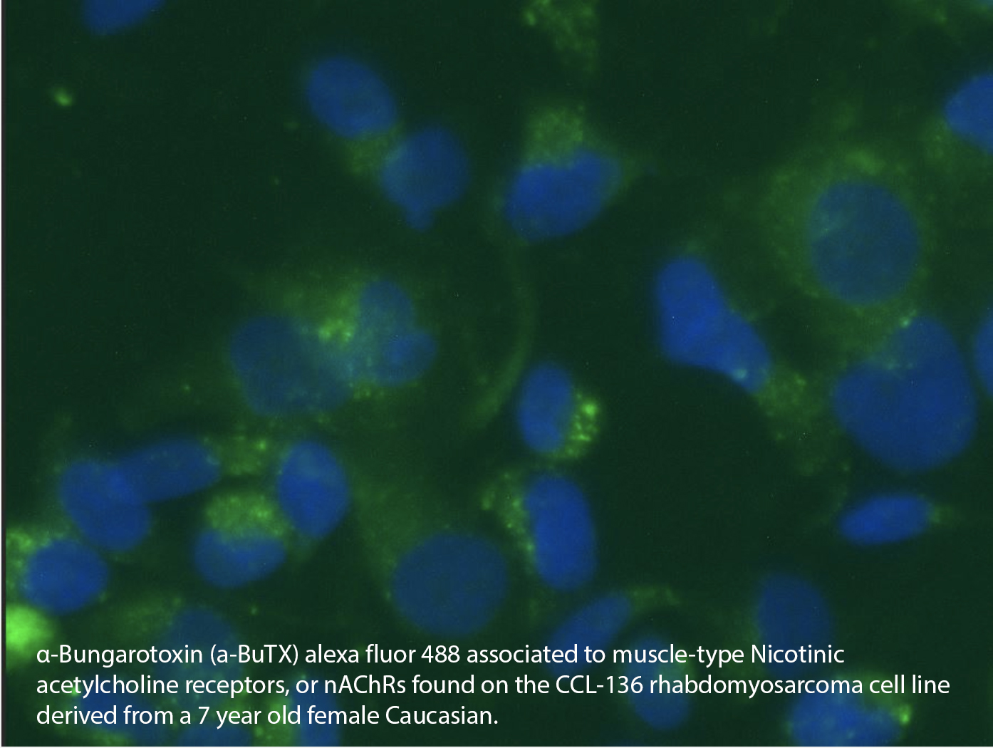
Cell membrane preparations containing nicotinic AChRs that are ligand-gated ion channels that form pores in cells plasma membranes, mediating fast signal transmission at synapses. Nicotinic AChRs are involved in a wide range of physiological processes, and can be either neuronal or muscle-type. The membrane preparations we have developed are suitable for receptor binding assays in which muscle type nicotinic AChRs are needed. The membranes are tested in several functional binding assays and quality testing criteria to meet binding specifications.
Other Products
[PS1000] FluoroStain™ Protein Fluorescent Staining Dye (Red, 1,000X), 1 ml
Product Info
Document
Product Info
Description
The FluoroStain™ Protein Fluorescent Staining Dye (Red, 1000×) is designed to substitute the common Coomassie Blue protein staining method, offering greater sensitivity and ease of operation. Unlike Coomassie Blue stain, the FluoroStain Protein Fluorescent Staining Dye binds to protein with high specificity, making destaining process an option rather than a requirement. With further reduction of background signals via destaining process, the FluoroStain™ is capable of achieving detection level parallel to silver staining without specialized imaging equipment, making it one of the most sensitive dyes available. In addition to its remarkable sensitivity, the FluoroStain™ Protein Fluorescent Staining Dye (Red, 1000×) brings a more reliable and safer user experience, since the stained gel can be visualized with blue-light illumination, avoiding the risk of skin/ eye damage caused by UV light. For best result, we suggest using B-BOX™ Blue Light LED epi-illuminator to visualize and analyze the gel stained with FluoroStain Protein Fluorescent Staining Dye (Red, 1000×). The FluoroStain™ Protein Fluorescent Staining Dye is compatible to the analysis of mass spectra, i.e. LC-MS/MS, MALDI-TOF, etc.
Spectral Characteristics
When it is bound with bovine serum albumin (BSA), the fluorescent emission of FluoroStain Protein Fluorescent Staining Dye can be excited by UV and blue light sources, with excitation peaks around 369 and 517 nm and emission at 605 nm. In absence of BSA, FluoroStain Protein Fluorescent Staining Dye shows ignorable fluorescence as compared with protein-bound form, therefore giving a clear background for photographic analysis.
These spectral characteristics made this fluorescent dye compatible with a wide variety of gel reading facilities, including UV/ blue light epi- and transilluminator, argon laser and mercury-arc lamp excitation gel scanners.
Storage
Protected from light -20°C for 24 months
Document
The FluoroStain™ Protein Fluorescent Staining Dye (Red, 1000×) is designed to substitute the common Coomassie Blue protein staining method, offering greater sensitivity and ease of operation. Unlike Coomassie Blue stain, the FluoroStain Protein Fluorescent Staining Dye binds to protein with high specificity, making destaining process an option rather than a requirement. With further reduction of background signals via destaining process, the FluoroStain™ is capable of achieving detection level parallel to silver staining without specialized imaging equipment, making it one of the most sensitive dyes available. In addition to its remarkable sensitivity, the FluoroStain™ Protein Fluorescent Staining Dye (Red, 1000×) brings a more reliable and safer user experience, since the stained gel can be visualized with blue-light illumination, avoiding the risk of skin/ eye damage caused by UV light. For best result, we suggest using B-BOX™ Blue Light LED epi-illuminator to visualize and analyze the gel stained with FluoroStain Protein Fluorescent Staining Dye (Red, 1000×). The FluoroStain™ Protein Fluorescent Staining Dye is compatible to the analysis of mass spectra, i.e. LC-MS/MS, MALDI-TOF, etc.
The SMO-HiFi™ DNA Polymerase is a new genetically modified, recombinant DNA polymerase with fidelity 70 times higher than Taq DNA polymerase during amplification, as well as very high elongation rate. Being highly thermostable, SMO-HiFi™ DNA Polymerase can remain viable even after being subjected to boiling for 2 minutes. The SMO-HiFi™ DNA Polymerase is also designed to operate in much lower Mg2+ concentration as compared to other DNA polymerase products.
Features
5’→3′ DNA polymerase activity
3’→5′ exonuclease (proofreading) activity
High reaction rate (up to 1 kb/10 seconds)
High fidelity, 70 times higher than Taq DNA polymerase
Blunt end amplicons
Thermo-stable: half-life is more than 10 hrs at 95°C
Storage
[TF1000] SMO-HiFi™ DNA Polymerase
-20°C for 24 months
Document
The SMO-HiFi™ DNA Polymerase is a new genetically modified, recombinant DNA polymerase with fidelity 70 times higher than Taq DNA polymerase during amplification, as well as very high elongation rate. Being highly thermostable, SMO-HiFi™ DNA Polymerase can remain viable even after being subjected to boiling for 2 minutes. The SMO-HiFi™ DNA Polymerase is also designed to operate in much lower Mg2+ concentration as compared to other DNA polymerase products.