

Description
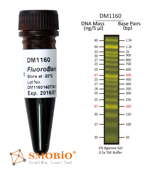
The DM1160 FluoroBand™ 50 bp Fluorescent DNA Ladder is a ready-to-use DNA ladder, which is pre-mixed with high sensitivity DNA binding fluorescent dye and loading dye for direct gel loading. The DNA Ladder DM1160 is composed of 17 individual DNA fragments: 1.5k, 1.2k, 1k, 900, 800, 700, 600, 500, 450, 400, 350, 300, 250, 200, 150, 100, and 50 bp derived from a mixture of PCR products and specifically digested plasmid DNA; these bands can be visualized when illuminated with 470 nm blue light or a UV light. This product contains two enhanced bands (500 bp and 200 bp) for easier reference. In addition, the low range Orange G tracking dye which mimics the migration of a 50 bp dsDNA during electrophoresis is also added for real time monitoring. Real time observation of the electrophoresis is also possible if compatible light source is fitted to the electrophoresis tank.
Features
- Sharp bands
- Quick reference— enhanced bands
- Ready-to-use— premixed with loading dye for direct loading
- Stable— room temperature storage over 6 months
- Directly observed by UV or blue light— premixed with high sensitive DNA fluorescent dye
Source
Phenol extracted PCR products and dsDNA digested with specific restriction enzymes, equilibrated in 10 mM Tris-HCl (pH 8.0) and 10 mM EDTA.
Range
50 ~ 1,500 bp
Concentration
54 µg/ 500 µl
Recommended loading volume
5 µl/ well
Storage
Protected from light
Room temperature for 6 months
4°C for 12 months
-20°C for 24 months


 • Detectable using a black light such as a black light UV flashlight or fluorescent lateral flow reader.