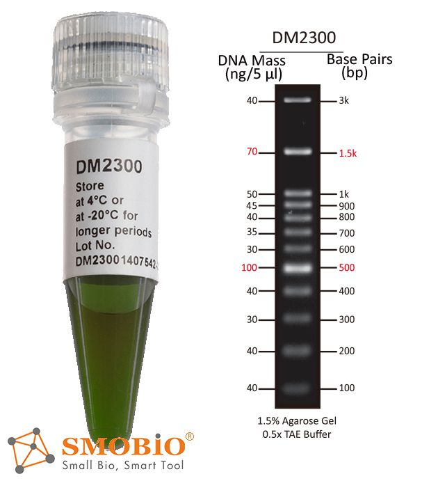
The DM2300 ExcelBand™ 100 bp+3K DNA Ladder is a ready-to-use DNA ladder, which is pre-mixed with loading dye for direct gel loading. The DNA ladder DM2300 is composed of 12 individual DNA fragments: 3k, 1.5k, 1k, 900, 800, 700, 600, 500, 400, 300, 200 and 100 bp derived from a mixture of PCR products and specifically digested plasmid DNA. This product contains two enhanced bands (1.5 kb and 500 bp) for easier reference. In addition, two tracking dyes, Xylene cyanol FF and Orange G which mimic the migration of 4,000 bp and 50 bp dsDNA during electrophoresis are also added for real time monitoring.
Detail
Description
The DM2300 ExcelBand™ 100 bp+3K DNA Ladder is a ready-to-use DNA ladder, which is pre-mixed with loading dye for direct gel loading. The DNA ladder DM2300 is composed of 12 individual DNA fragments: 3k, 1.5k, 1k, 900, 800, 700, 600, 500, 400, 300, 200 and 100 bp derived from a mixture of PCR products and specifically digested plasmid DNA. This product contains two enhanced bands (1.5 kb and 500 bp) for easier reference. In addition, two tracking dyes, Xylene cyanol FF and Orange G which mimic the migration of 4,000 bp and 50 bp dsDNA during electrophoresis are also added for real time monitoring.
Features
Sharp bands
Quick reference— enhanced bands
Ready-to-use— premixed with loading dye for direct loading
Stable— room temperature storage over 6 months
Source
Phenol extracted PCR products and dsDNA digested with specific restriction enzymes, equilibrated in 10 mM Tris-HCl (pH 8.0) and 10 mM EDTA.
Range
100 ~ 3,000 bp
Concentration
56 µg/ 500 µl
Recommended loading volume
5 µl/ well
Storage
Room temperature for 6 months 4°C for 12 months -20°C for 36 months
Other Products
APOPercentage apoptosis assay kit
Product Info
Document
Product Info
What is Apoptosis?
Apoptosis is an essentially normal physiological process that removes now redundant, cells, particularly during embryonic development and early growth. In adult animals the process removes cells that are irreparable. The apoptotic process is also involved in many major diseases such as cancer, where transformed tumour cells have their apoptotic process disabled, permitting cell cycling to continue unchecked. In contrast some forms of senile dementia may result from excessive apoptotic induction of neural cells.
The apoptotic process in mammalian cells is a rapid event (2‐4 hours). Within this short time span an apparently viable cell can be quietly dismantled, to disappear leaving no visible trace of its former existence.
How is apoptosis detected or measured?
An apoptosis cascade of activators, effectors and regulators has been identified. This in turn led to a range of apoptosis assays being devised to detect and monitor these events. Some laboratories will employ two distinct assays, one selected to detect early (initiation) apoptotic events, while a second assay will target a later (execution) event. Apoptosis assays, based on methodology, can be classified into four major inter‐linked groups:
[1] DNA fragmentation (electrophoresis and nick end labelling, TUNEL).
[2] Apoptotic proteases (fluorescently labelled antibodies to the caspases).
[3] Flow cytometric analysis (FACS, incorporating other group assays).
Biocolor’s APOPercentage assay is based on the latter. Further information can be found under the ‘Mode of Action’ Tab.
How does APOPercentage detect apoptosis?
The mammalian cell membrane has been described as a semi‐fluid mosaic structure, composed of phospholipids with a diverse group of inserted proteins and some cholesterol. The phospholipids are the major components of the membrane and are arranged in the form of a ‘bi‐layer’; which is asymmetric in composition, structure, and function.
To ensure normal transmembrane functions the phospholipids must be maintained in an asymmetric composition. The process is regulated by ‘flippases’, which catalyse the active transport of aminophospholipids from the outer to inner monolayer. However, in cells undergoing apoptosis, flippase is overwhelmed by the action of another enzyme, termed ‘floppase’ or ‘scramblase’. The net effect is a scrambling of the phospholipid distribution between the inner and outer monolayers.
Cell membrane changes during apoptosis
The APOPercentage assay utilises an intense, pink-coloured dye reagent which is taken up during in-vitro culture by apoptosis-committed cells. This uptake occurs at the stage of Phosphatidylserine transmembrane movement, as produced by the flipflop mechanism. Dye uptake continues until blebbing occurs. No further dye can then enter the now defunct cell and the dye that has accumulated within the cell is not released (unlike necrotic cells which release dye).
Since the dye reagent is excluded or not retained by healthy or necrotic cells it therefore acts as a specific label for apoptotic cells.
How are APOPercentage-labelled cells quantified?
Labelled apoptosis cells may then by conveniently analysed by the following methods:
Direct Analysis The intense pink colour of the labelled cells can be visually assessed using brightfield microscopy. Apoptosis in substrate-adherent cell populations is therefore readily quantified using image analysis techniques. This technique is the most sensitive with the ability of detecting one single apoptotic cell per well.
Colorimetry protocol Dye that accumulates within apoptotic cells is released into solution via addition of Dye Release Reagent. The concentration of this intracellular dye is then measured at 550nm using a microplate colorimeter/spectrophotometer.
NB: The APOPercentage assay kit does NOT require the use of a Flow Cytometer.
Limit of Detection
A single cell (via image analysis method)
Detection Method
Colorimetric (550nm) (Endpoint) or Image Analysis based
Measurements per kit
Sufficient for 4×24 well plates or 6×96 well plates
Suitable Samples
Adherent mammalian cells (in-vitro)
APOPercentage kit contents:
1. APOPercentage Dye (1x5ml)
2. Dye Release Reagent (1x150ml)
3. Phosphate Buffered Saline (PBS) (1x120ml)
4. 24-well starter plate.
5. Assay kit manual.
The Colorimetric Protocol requires a Microplate Colorimeter / Spectrophotometer.
Additional 96-well plates will be required for use when reading dye absorbance values.
The Direct Detection Protocol Requires an inverted stage microscope with an attached digital camera.
NB: Additional reagents (typically culture medium and suitable apoptosis treatments) may be required for sample preparation prior to assay. Consult manual or contact us for further details.
Document
The APOPercentage™ Apoptosis kit is a dye-based, colorimetric assay for detection and measurement of apoptosis (programmed cell death) during in-vitro cell culture.
96-Well Low Elution Ring Magnetic Plate With Integrated Cushion Base
Product Info
Document
Product Info
The Permagen ring magnet low elution plate with integrated cushion base plate was designed for use in automation applications where volumes as low as 5 µL are required. Most PCR plates are bent, leading to inconsistent lab results. Unlike most products, we have added an angled frame around the top of our plate, this helps with two things, straightening out the PCR plate, and leading it to the proper location on the magnets during automation while the cushion assist compensates for labware inconsistencies.
SBS SLAS Footprint (127.75mm x 85.50mm) to fit into any automated liquid handling robot
Features include solid aluminum alloy construction and hard coat anodized finish for years of trouble-free use, and compatible with any magnetic beads
The Permagen ring magnet low elution plate with integrated cushion base plate was designed for use in automation applications where volumes as low as 5 µL are required. Most PCR plates are bent, leading to inconsistent lab results. Unlike most products, we have added an angled frame around the top of our plate, this helps with two things, straightening out the PCR plate, and leading it to the proper location on the magnets during automation while the cushion assist compensates for labware inconsistencies.
CE-IVD marked version available for in vitro diagnostic use
Available in TaqMan format for analysis.
Giardiasis is a disease of the small bowel caused by the protozoan parasite Giardia intestinalis (syn.duodenalis or lamblia). Giardia is one of the most common intestinal parasites in the world, and occurs at very high prevalence rates in places with poor water sanitation. Individuals become infected through ingesting or coming into contact with contaminated water, food or soil. It can also be spread through the faecal-oral route due to poor hygiene practices, which makes it common in day-care centers. G. intestinalis lives inside the intestines of infected humans or other animals including cats, dogs, birds, cows, beaver and deer. Symptoms of infection include diarrhea, malaise, excessive gas, bloating, nausea, diminished interest in food, possible vomiting and weight loss.
There are 2 kits available for the detection of Giardia intestinalis:
Storage Conditions and Product Stability All kit components can be stored for 1 year after the date of production without showing any reduction in performance.
All kit components should be stored at -20°C upon arrival. Repeated thawing and freezing (> 2 x) of the Master Mix and Positive Control should be avoided, as this may affect the performance of the assay. If the reagents are to be used only intermittently, they should be frozen in aliquots.