[DS1000] FluoroStain™ DNA Fluorescent Staining Dye (Green, 10,000X), 500 μl
Facebook
X
Pinterest
Email
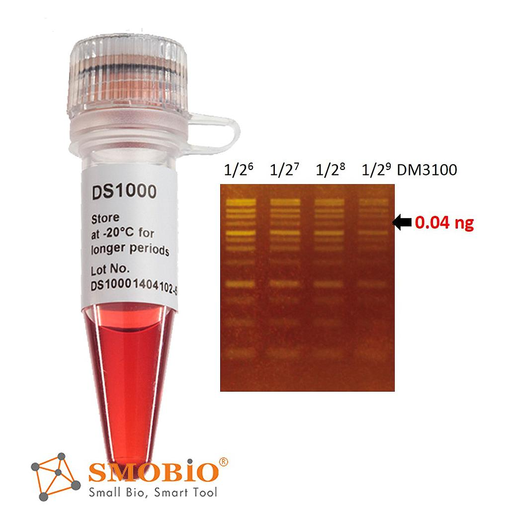
The FluoroStain™ DNA Fluorescent Staining Dye is designed to be a safer replacement for conventional Ethidium bromide (EtBr) which poses a significant health and safety hazard for its users. The FluoroStain™ DNA Fluorescent Staining Dye offers at least 10 times sensitivity in DNA detection levels, and is capable of detecting double stranded DNA (dsDNA) fragments up to 0.04 ng in electrophoresis analysis. The FluoroStain™ DNA Fluorescent Staining Dye shows a high specificity to the dsDNA, with negligible background signal, making the destaining process entirely optional. FluoroStain™ DNA Fluorescent Staining Dye is compatible with both the conventional ultra violet gel-illuminating systems as well as the less harmful long wave length blue light illumination systems. The emission when bound to dsDNA is 522 nm, while its excitation peaks are at 270, 370 and 497 nm.
Detail
Description
The FluoroStain™ DNA Fluorescent Staining Dye is designed to be a safer replacement for conventional Ethidium bromide (EtBr) which poses a significant health and safety hazard for its users. The FluoroStain™ DNA Fluorescent Staining Dye offers at least 10 times sensitivity in DNA detection levels, and is capable of detecting double stranded DNA (dsDNA) fragments up to 0.04 ng in electrophoresis analysis. The FluoroStain™ DNA Fluorescent Staining Dye shows a high specificity to the dsDNA, with negligible background signal, making the destaining process entirely optional. FluoroStain™ DNA Fluorescent Staining Dye is compatible with both the conventional ultra violet gel-illuminating systems as well as the less harmful long wave length blue light illumination systems. The emission when bound to dsDNA is 522 nm, while its excitation peaks are at 270, 370 and 497 nm.
Features:
Excellent for post staining
Sensitivity: 0.04 ng DNA
A safer alternative to EtBr
Compatibility: suitable to blue or UV light
Increased cloning efficiency (blue light)
Storage
Protected from light 4°C for 12 months -20°C for 24 months
Other Products
PACE® TRIAL KITS
Product Info
Document
Product Info
ABOUT
Everything you need to run a trial PACE® allele-specific PCR Genotyping Reaction on your existing lab equipment. Each PACE Trial Kit includes Test DNA samples, PACE Genotyping Assays, PACE Master Mix and a comprehensive PACE Genotyping Trial Kit Manual.
WHO IS THIS TRIAL KIT FOR?
Anyone who wants to try PACE genotyping reagents in their lab for the first time with a set of validated DNA samples, SNP assays and PACE Master Mix.
TRIAL KIT OVERVIEW
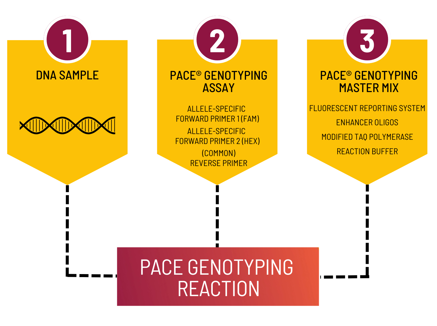
Step 1. Dispense each of the three trial DNA samples (DNA 1, 2 and 3) plus water (No Template Control) in triplicate onto a PCR plate using the suggested volumes.
Step 2. Combine appropriate volumes of PACE Genotyping Master Mix with PACE Genotyping Assay in a tube, as directed, then mix.
Step 3. Dispense the combined mixtures into each of the wells containing DNA using volumes indicated. Each test now contains a complete PACE Genotyping Reaction.
Step 4. Seal your PCR plate with an optically clear seal and centrifuge to ensure all components are at the bottom of the wells.
Step 5.Thermally cycle the reaction plate using the thermal cycling conditions provided.
Step 6. Read the plate and compare data produced with the expected results provided in the manual. Simple!
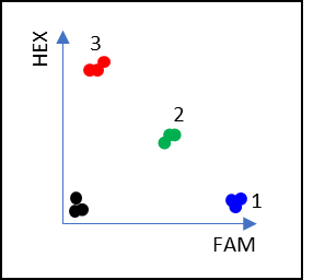
PACE MECHANISM
More information on the PACE genotyping chemistry and how it works can be found here: www.3crbio.com/#pace. PACE allele-specific PCR is used for the detection of SNPs, Indels and other sequence variants.
REQUIRED COMPONENTS
qPCR machine or Thermocycler + Fluorescent plate reader
PCR plate or equivalent and appropriate optically clear seal
The HiPure Liquid RNA & miRNA Kit integrates phenol/guanidine-based sample lysis and silica membrane purification of total RNA. MagZol LS Reagent, included in the kits, is a monophasic solution of phenol and guanidine thiocyanate designed to facilitate lysis of liquid sample and inhibit RNase. The high lysis efficiency of the reagent and the subsequent removal of contaminants by organic phase extraction enable the use of up to 0.25ml liquid sample per mini spin column.
Details
Specifications
Features
Specifications
Main Functions
Isolation total RNA from 0.25ml body fluids using column and MagZol LS reagent
Applications
qPCR / RT-PCR, liquid or solid-phasechip analysis, hybridization and SNP detection
0.25ml liquid samples are homogenized in 0.75ml MagZol LS Reagent. After addition of chloroform, the homogenate is separated into aqueous and organic phases by centrifugation. The upper, aqueous phase is extracted, and ethanol is added to provide appropriate binding conditions. The sample is then applied to the spin column, where the total RNA (up to 100 µg) binds to the membrane and phenol and other contaminants are efficiently washed away. High-quality RNA is then eluted in 30–100µl of RNase-free water.
Advantages
High quality – high purity total RNA can be directly used in various sensitive downstream applications
Fast – isolation of several samples can be completed in 40 minutes by using column purification method
Safety – no phenol chloroform extraction required
Sensitive – direct lysis of blood, plasma and other samples without separation of leukocytes
Kit Contents
Contents
R416302
R416303
Purification Times
50 Preps
250 Preps
HiPure RNA Mini Columns
50
250
2ml Collection Tubes
50
2 x 125
MagZol LS Reagent
60 ml
270 ml
Buffer RWC
20 ml
60 ml
Buffer RW2*
20 ml
2 x 50 ml
RNase Free Water
10 ml
30 ml
Storage and Stability
MagZol LS Reagent should be stored at 2-8°C upon arrival. However, short-term storage (up to 24 weeks) at room temperature (15-25°C) does not affect their performance. The remaining kit components can be stored dry at room temperature (15-25°C) and are stable for at least 18 months under these conditions.
Document
The HiPure Liquid RNA & miRNA Kit integrates phenol/guanidine-based sample lysis and silica membrane purification of total RNA. MagZol LS Reagent, included in the kits, is a monophasic solution of phenol and guanidine thiocyanate designed to facilitate lysis of liquid sample and inhibit RNase. The high lysis efficiency of the reagent and the subsequent removal of contaminants by organic phase extraction enable the use of up to 0.25ml liquid sample per mini spin column.
With the development of molecular biology, stool, a new non-invasive sample, has been widely used in the research of animal molecular genetics, population ecology, behavioral ecology and some intestinal disease diagnosis. Stool samples includes gut microbial DNA, food residue sample DNA, and alimentary tract exfoliated cell DNA.
The primary problem encountered when using stool sample for molecular biology research is the low content of exfoliated cells in the digestive tract and a certain degree of degradation of genetic material in stool. Another issue in molecular scatology research based on PCR is the presence of a large number of inhibitors in stool that can affect Taq enzyme activity, leading to downstream detection inactivation. These inhibitors include polysaccharides, plant polysaccharides, bile acids, bile salts, bile pigments, digestive juices, mucus, etc. Therefore, selecting appropriate extraction methods to obtain high-quality DNA is the key to successful downstream detection of stool DNA.
At present, the pretreatment methods used in the laboratory, such as phenol/chloroform extraction, cetyltrimethyl bromide (CTAB) lysis, and guanidine isothiocyanate lysis, lack universality in different species, and the success rate of extracting DNA for PCR amplification is also very low. The HiPure Stool DNA Kit provided by Magen Company has opened up a new approach for DNA extraction from stool samples with good universality, high cost-effectiveness, high yield and purification. The reagent kit adopts a unique solution system and inhibitory factor adsorbent, which can efficiently remove various impurities in stool samples. The purified DNA can be directly used for PCR, quantitative PCR and other applications.
This product allows rapid and reliable isolation of high-quality genomic DNA from various stool samples. Up to 100 mg soil samples can be processed in 60 minute. The system combines the reversible nucleic acid binding properties of HiPure matrix with the speed and versatility of spin column technology to eliminate PCR inhibiting compounds such as humic acid from soil samples. Purified DNA is suitable for PCR, restriction digestion, and next-generation sequencing. There are no organic extractions thus reducing plastic waste and hands-on time to allow multiple samples to be processed in parallel.
Details
Specifications
Features
Specifications
Main Functions
Isolation total DNA from 50-100mg stool samples
Applications
PCR, Southern Blot, enzyme digestion and NGS, etc.
Purification method
Mini spin column
Purification technology
Silica technology
Process method
Manual (centrifugation or vacuum)
Sample type
Stool
Sample amount
50-100mg
Yield
3-15μg
Elution volume
≥30μl
Time per run
≤60 minutes
Liquid carrying volume per column
750μl
Binding yield of column
100μg
Principle
Stool sample is homogenized and then treated in a specially formulated buffer containing detergent to lyse bacteria, yeast, and fungal samples. Humic acid, proteins, polysaccharides, and other contaminants are removed using our proprietary Absorber Solution. Binding conditions are then adjusted and the sample is applied to a DNA Mini Column. Two rapid wash steps remove trace contaminants and pure DNA is eluted in low ionic strength buffer. Purified DNA can be directly used in downstream applications without the need for further purification.
Advantages
High purity – unique adsorbent can completely remove inhibitory factors
High concentration – maximum extraction of total DNA from stool samples
High recovery – DNA can be recovered at the level of PG
Good repeatability – silica technology can obtain ideal results every time
Kit Contents
Contents
D314102
D314103
Purification Times
50 Preps
250 Preps
HiPure DNA Mini Columns II
50
250
2ml Collection Tubes
50
250
2ml Bead Tubes
50
250
Proteinase K
24 mg
120 mg
Protease Dissolve Buffer
1.8 ml
10 ml
Buffer SPL
40 ml
200 ml
Buffer PCI
40 ml
200 ml
Buffer AL
20 ml
80 ml
Buffer GW1
22 ml
88 ml
Buffer GW2
20 ml
2 x 50 ml
Buffer AE
15 ml
30 ml
Storage and Stability
Proteinase K and Buffer PCI should be stored at 2-8°C upon arrival. However, short-term storage (up to 12 weeks) at room temperature (15-25°C) does not affect their performance. The remaining kit components can be stored at room temperature (15-25°C) and are stable for at least 18 months under these conditions. The entire kit can be stored at 2–8°C, but in this case buffers should be redissolved before use. Make sure that all buffers are at room temperature when used.
Document
With the development of molecular biology, stool, a new non-invasive sample, has been widely used in the research of animal molecular genetics, population ecology, behavioral ecology and some intestinal disease diagnosis. Stool samples includes gut microbial DNA, food residue sample DNA, and alimentary tract exfoliated cell DNA.
The primary problem encountered when using stool sample for molecular biology research is the low content of exfoliated cells in the digestive tract and a certain degree of degradation of genetic material in stool. Another issue in molecular scatology research based on PCR is the presence of a large number of inhibitors in stool that can affect Taq enzyme activity, leading to downstream detection inactivation. These inhibitors include polysaccharides, plant polysaccharides, bile acids, bile salts, bile pigments, digestive juices, mucus, etc. Therefore, selecting appropriate extraction methods to obtain high-quality DNA is the key to successful downstream detection of stool DNA.
At present, the pretreatment methods used in the laboratory, such as phenol/chloroform extraction, cetyltrimethyl bromide (CTAB) lysis, and guanidine isothiocyanate lysis, lack universality in different species, and the success rate of extracting DNA for PCR amplification is also very low. The HiPure Stool DNA Kit provided by Magen Company has opened up a new approach for DNA extraction from stool samples with good universality, high cost-effectiveness, high yield and purification. The reagent kit adopts a unique solution system and inhibitory factor adsorbent, which can efficiently remove various impurities in stool samples. The purified DNA can be directly used for PCR, quantitative PCR and other applications.
This product allows rapid and reliable isolation of high-quality genomic DNA from various stool samples. Up to 100 mg soil samples can be processed in 60 minute. The system combines the reversible nucleic acid binding properties of HiPure matrix with the speed and versatility of spin column technology to eliminate PCR inhibiting compounds such as humic acid from soil samples. Purified DNA is suitable for PCR, restriction digestion, and next-generation sequencing. There are no organic extractions thus reducing plastic waste and hands-on time to allow multiple samples to be processed in parallel.