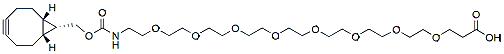
endo-BCN-PEG8-acid is a BCN-containing crosslinker reagent. The terminal carboxylic acid is reactive with primary amine groups in the presence of activators (e.g. EDC, or HATU). The BCN group enable copper free click chemistry with azide -tagged biomolecules. The hydrophilic PEG spacer increases solubility in aqueous media. Reagent grade, for research purpose. Please contact us for GMP-grade inquiries.
Detail
endo-BCN-PEG8-acid is a BCN-containing crosslinker reagent. The terminal carboxylic acid is reactive with primary amine groups in the presence of activators (e.g. EDC, or HATU). The BCN group enable copper free click chemistry with azide -tagged biomolecules. The hydrophilic PEG spacer increases solubility in aqueous media. Reagent grade, for research purpose. Please contact us for GMP-grade inquiries.
Aflatoxin is the most common food toxin that is harmful to human and animal health. The most frequent aflatoxins are B1, B2, G1, and G2, which can affect the body through respiratory, mucosal, or cutaneous routes, causing an excessive inflammatory response. Aflatoxin can infect crops during their growing stages or even after they are harvested. It mainly targets the liver and can impair the effectiveness of immunization in children, increasing the risk of infection. Aflatoxin detection and quantification in food and feed is a critical part of food and feed safety concerns.
Document
Aflatoxin is the most common food toxin that is harmful to human and animal health. The most frequent aflatoxins are B1, B2, G1, and G2, which can affect the body through respiratory, mucosal, or cutaneous routes, causing an excessive inflammatory response. Aflatoxin can infect crops during their growing stages or even after they are harvested. It mainly targets the liver and can impair the effectiveness of immunization in children, increasing the risk of infection. Aflatoxin detection and quantification in food and feed is a critical part of food and feed safety concerns.
Storage term: stored at ≤-20℃,keep away from light, avoid heavy weight and repeated freezing and thawing.
Term of Validity: 14 months
Isothermal nucleic acid Principle Summary
This kit is based on a rapid nucleic acid amplification technology at room temperature and constant temperature: at room temperature and constant temperature (generally 39 ºC~42 ºC), reverse transcriptase uses specific primers and template RNA to synthesize cDNA strands, and binds the auxiliary protein and single strand With the help of the protein, the recombinase and the primer form a complex; perform a homology search and bind the target homology domain, at this time a D-loop region is formed at the homology position and strand exchange begins; accompanied by the recombinase from the complex Upon dissociation, the polymerase also binds to the 3′ end of the primer, initiating chain extension. Relying on the action of nfo enzyme, adding specific molecular probes designed according to the template, and using colloidal gold technology (sandwich method) can detect the final result.
Isothermal nucleic acid Product Features
1/ High sensitivity and specificity, short reaction time.
2/ The reagent form is freeze-dried, stable and easy to operate.
3/ The reaction can be operated by metal bath and water bath pot without purchasing expensive PCR apparatus.
Technical Parameters:
Parameters
Details
Product Name
RNA Isothermal Amplification Kit NFO
Manufacturer
Amp-future
Storage Temperature
-20°C
Kit Components
Enzymes, Buffers ,Reagents
Packaging
48 Tests/box
Detection Limit
500-1000copies/µL
Shipping
ICE
Test Time
5-20mins
Isothermal nucleic acid Applications
Suitable for RNA isothermal rapid amplification kit(NFO type)
Primer: Require pair of nucleotide primers with the length of 25-35 bp.
RNA NFO kit reaction temperature is 39 to 42℃ and time is 5-20 minutes.
Notes
1/ Please avoid nucleic acid contamination and set blank control during reaction due to the high sensitivity of the kit.
2/ Please take out the required quantity of MIRA reaction units for the experiment, and put the rest under storage conditions when performing the experiment.