This product has been discontinued (see here), please use the recommended replacement product β-Glucan Assay Kit (Yeast and Mushroom) for all your yeast and mushroom β-Glucan testing needs.
Detail
K-EBHLG
SKU: 700004278
50 assays per kit
Content:
50 assays per kit
Shipping Temperature:
Ambient
Storage Temperature:
Short term stability: 2-8oC, Long term stability: See individual component labels
Stability:
> 2 years under recommended storage conditions
Analyte:
β-Glucan
Assay Format:
Spectrophotometer
Detection Method:
Absorbance
Wavelength (nm):
510
Signal Response:
Increase
Limit of Detection:
1 g/100 g
Reaction Time (min):
~ 100 min
Application examples:
Yeast preparations and other materials
Method recognition:
Novel method
This product has been discontinued (see here), please use the recommended replacement product β-Glucan Assay Kit (Yeast and Mushroom) for all your yeast and mushroom β-Glucan testing needs.
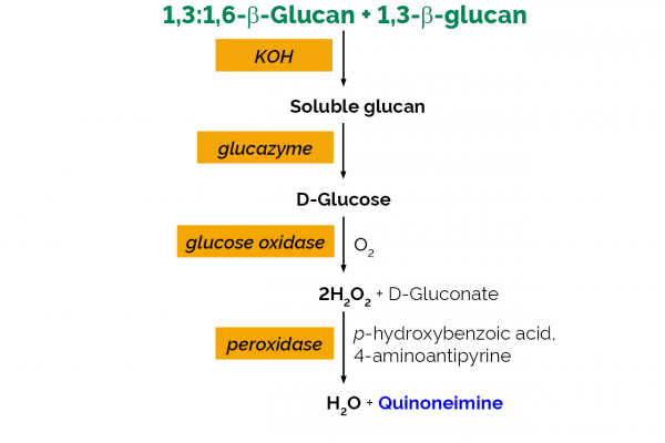
Enzymatic Yeast Beta-Glucan test kit, an enzymatic procedure for the measurement and analysis of 1,3:1,6-β-glucan in yeast. Also measures 1,3-β-glucan.
All reagents stable for > 12 months after preparation
Mega-Calc™ software tool is available from our website for hassle-free raw data processing
Other Products
APOPercentage apoptosis assay kit
Product Info
Document
Product Info
What is Apoptosis?
Apoptosis is an essentially normal physiological process that removes now redundant, cells, particularly during embryonic development and early growth. In adult animals the process removes cells that are irreparable. The apoptotic process is also involved in many major diseases such as cancer, where transformed tumour cells have their apoptotic process disabled, permitting cell cycling to continue unchecked. In contrast some forms of senile dementia may result from excessive apoptotic induction of neural cells.
The apoptotic process in mammalian cells is a rapid event (2‐4 hours). Within this short time span an apparently viable cell can be quietly dismantled, to disappear leaving no visible trace of its former existence.
How is apoptosis detected or measured?
An apoptosis cascade of activators, effectors and regulators has been identified. This in turn led to a range of apoptosis assays being devised to detect and monitor these events. Some laboratories will employ two distinct assays, one selected to detect early (initiation) apoptotic events, while a second assay will target a later (execution) event. Apoptosis assays, based on methodology, can be classified into four major inter‐linked groups:
[1] DNA fragmentation (electrophoresis and nick end labelling, TUNEL).
[2] Apoptotic proteases (fluorescently labelled antibodies to the caspases).
[3] Flow cytometric analysis (FACS, incorporating other group assays).
Biocolor’s APOPercentage assay is based on the latter. Further information can be found under the ‘Mode of Action’ Tab.
How does APOPercentage detect apoptosis?
The mammalian cell membrane has been described as a semi‐fluid mosaic structure, composed of phospholipids with a diverse group of inserted proteins and some cholesterol. The phospholipids are the major components of the membrane and are arranged in the form of a ‘bi‐layer’; which is asymmetric in composition, structure, and function.
To ensure normal transmembrane functions the phospholipids must be maintained in an asymmetric composition. The process is regulated by ‘flippases’, which catalyse the active transport of aminophospholipids from the outer to inner monolayer. However, in cells undergoing apoptosis, flippase is overwhelmed by the action of another enzyme, termed ‘floppase’ or ‘scramblase’. The net effect is a scrambling of the phospholipid distribution between the inner and outer monolayers.
Cell membrane changes during apoptosis
The APOPercentage assay utilises an intense, pink-coloured dye reagent which is taken up during in-vitro culture by apoptosis-committed cells. This uptake occurs at the stage of Phosphatidylserine transmembrane movement, as produced by the flipflop mechanism. Dye uptake continues until blebbing occurs. No further dye can then enter the now defunct cell and the dye that has accumulated within the cell is not released (unlike necrotic cells which release dye).
Since the dye reagent is excluded or not retained by healthy or necrotic cells it therefore acts as a specific label for apoptotic cells.
How are APOPercentage-labelled cells quantified?
Labelled apoptosis cells may then by conveniently analysed by the following methods:
Direct Analysis The intense pink colour of the labelled cells can be visually assessed using brightfield microscopy. Apoptosis in substrate-adherent cell populations is therefore readily quantified using image analysis techniques. This technique is the most sensitive with the ability of detecting one single apoptotic cell per well.
Colorimetry protocol Dye that accumulates within apoptotic cells is released into solution via addition of Dye Release Reagent. The concentration of this intracellular dye is then measured at 550nm using a microplate colorimeter/spectrophotometer.
NB: The APOPercentage assay kit does NOT require the use of a Flow Cytometer.
Limit of Detection
A single cell (via image analysis method)
Detection Method
Colorimetric (550nm) (Endpoint) or Image Analysis based
Measurements per kit
Sufficient for 4×24 well plates or 6×96 well plates
Suitable Samples
Adherent mammalian cells (in-vitro)
APOPercentage kit contents:
1. APOPercentage Dye (1x5ml)
2. Dye Release Reagent (1x150ml)
3. Phosphate Buffered Saline (PBS) (1x120ml)
4. 24-well starter plate.
5. Assay kit manual.
The Colorimetric Protocol requires a Microplate Colorimeter / Spectrophotometer.
Additional 96-well plates will be required for use when reading dye absorbance values.
The Direct Detection Protocol Requires an inverted stage microscope with an attached digital camera.
NB: Additional reagents (typically culture medium and suitable apoptosis treatments) may be required for sample preparation prior to assay. Consult manual or contact us for further details.
Document
The APOPercentage™ Apoptosis kit is a dye-based, colorimetric assay for detection and measurement of apoptosis (programmed cell death) during in-vitro cell culture.
For simplified collection and shipment of leaf samples for our premium DNA Extraction and Genotyping Lab Services.
About
3CR Bio’s Leaf Sample Collection Kit – Your Gateway to High-Quality DNA Extraction and Genotyping Lab Services
The correct sampling and storage of leaf samples for DNA extraction have a significant impact on the final quality and quantity of the DNA extracted. Our simple, lightweight kits provide everything you need to sample and ship your leaf tissue from anywhere in the world, so it reaches us in top condition.
Use the 3CR Bioscience Leaf Sample Collection Kit to sample and prepare leaf tissue optimally when shipping to us for DNA extraction and Genotyping. Full instructions available in the Leaf Sample Collection User Guide.
Kit Contents
1 x 96-tube storage rack containing individual 2D-barcoded tubes sealed with a secure locking lid.
1 x Sponge pad to fit inside the lid of the storage rack for shipping.
1 x Sealed black plastic packet containing 2 x 10g sachets of desiccant.
1 x Large clear sealable plastic bag.
1 x Basic leaf cutting tool and leaf cutting mat (order separately)
or
1 x Advanced cutting tool (order separately)
Plate Map for recording sample IDs (image not shown, provided in paper and electronic form)
The Plant Kit is used in combination with our Lab Services. Visit the Lab Services page for more information about our DNA Extraction, Endpoint Genotyping, and GBTS services.
Here you can also:
View an interactive workflow infographic detailing our user-friendly ordering, shipping, processing, and data retrieval.
Access our online customer portal to register, submit projects. and review data.
Solid Phase Adsorption Toxin Tracking (SPATT) is a biomimetic in-situ water monitoring tool that falls under an expanding umbrella of passive samplers. It serves to warn researchers of toxin-producing harmful algal bloom (HAB) developments early on. It has been popularized through its affordability, ease of use, and its ability to capture ephemeral events in marine, brackish, and freshwater environments. Its uptake of contaminants has been shown to be more similar than other sampling methods to that of aquatic species like bivalves, mussels, and clams. It provides an average bioavailable fraction of a toxin over deployment time that can be used to determine an overall toxin risk to organisms. The sampling period typically depends on the bioactivity at a site, ranging from 24 hours to 4 weeks in most cases.
A SPATT passively absorbs and desorbs extracellular compounds over its stretch of time at a sampling site; in an organism, a toxin would go through biochemical detoxification processes. Passive samplers have a higher sensitivity for more compounds and provide improved stability and preservation of these compounds within the resin. SPATT devices capture less commonly detected cyanotoxins (e.g. cylindrospermopsin) at lower concentrations than that of a grab sample (collected at one point in time). Grab samples are limited in scope and sensitivity, and underrepresent toxins like microcystin-LR, which is picked up very reliably through SPATT technology.
Uses HP20 that is widely applicable for many toxins.
Used to capture:
Cyanotoxin (e.g. microcystin and cylindrospermopsin)
Saxitoxin & derivatives (GNTXs, C-toxins), and other paralytic shellfish toxins (PSTs)