
Description
Specifications
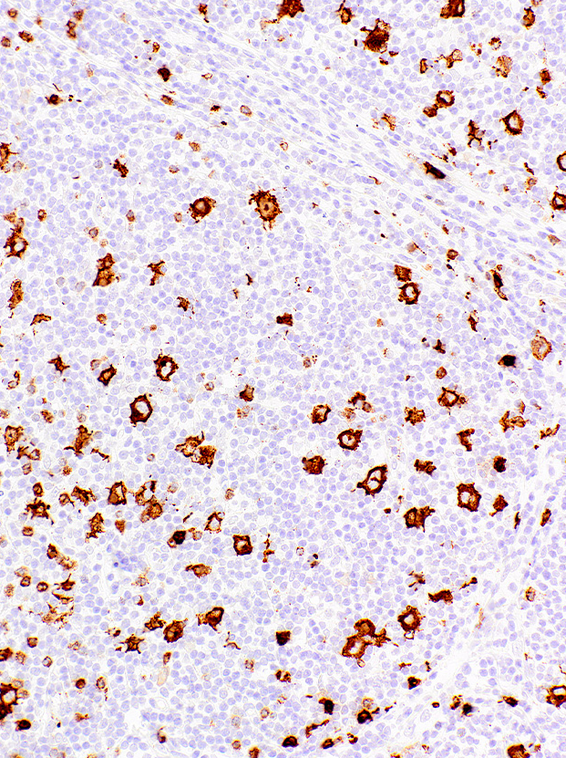
| Clone | IHC527 |
| Source | Mouse Monoclonal |
| Positive Control | Hodgkin’s Lymphoma |
| Dilution Range | 1:200 |

| Clone | IHC527 |
| Source | Mouse Monoclonal |
| Positive Control | Hodgkin’s Lymphoma |
| Dilution Range | 1:200 |

N-(t-butyl ester-PEG4)-N-bis(PEG4-Propargyl) is a trifunctional PEG linker that combines a t-butyl ester with two terminal alkynes. The alkynes can be applied in copper-click chemistry with azides to form stable triazole linkages with a target molecule, while the t-butyl ester can be deprotected and reacted with amines or alcohols to form amides or esters.

N-(t-butyl ester-PEG4)-N-bis(PEG4-Propargyl) is a trifunctional PEG linker that combines a t-butyl ester with two terminal alkynes. The alkynes can be applied in copper-click chemistry with azides to form stable triazole linkages with a target molecule, while the t-butyl ester can be deprotected and reacted with amines or alcohols to form amides or esters.

Propargyl-PEG5-acid has a propargyl group at one end and an acid group at the other end. The acid can react with primary amines to form a stable amide bond, activation will be needed. The PEG units enhances solubility of the molecule in aqueous environment. The propargyl group can be linked to azide-containing biomolecules via Click Chemistry. Reagent grade, for research purpose. Please contact us for GMP-grade inquiries.

Propargyl-PEG5-acid has a propargyl group at one end and an acid group at the other end. The acid can react with primary amines to form a stable amide bond, activation will be needed. The PEG units enhances solubility of the molecule in aqueous environment. The propargyl group can be linked to azide-containing biomolecules via Click Chemistry. Reagent grade, for research purpose. Please contact us for GMP-grade inquiries.

Product Highlights
Speed: Adjustable speed, up to 4500 rpm
Vortex Oscillation Intensity:1-4 intensity levels, selectable and adjustable
Programming:Programmable,freely choose combinations of oscillation strength,duration.centrifugation speed, and time
Safety:Safety interlock device to ensure user safety
Compatibility: Single tube or multi-tube centrifugation/mixing with one-button operation, saving time and cost
Display:Touch buttons and an LCD screen clearly indicate and set oscillation level, duration, and cycle count
Convenience: Multi-functional, compact design, occupies minimal space
Technical Specifications
| Dimensions (mm) | 200(w)x243(H)x127(D) | Working temperature | 5℃ to 40℃ |
| Oscillation Duration | 0-30 seconds (increment of 1 second) | TD4Z Speed range | 1000-3600 rpm (increment of 100 rpm) |
| Power Supply | AC 220V 50/60 Hz | Spin Timer | 0 seconds to 30 minutes |
| Weight | 3.5kg (including power supply) | Oscillation Intensity | Level 1-4 |
| Speed range TD4Z | 1-99 programs | Centrifugation Adjustment TD4Z | 1000-3600 rpm (increment of 100 rpm) |
PCR laboratories and general molecular biology laboratories
Model:
TD4Z
Brand:
Yingtai
Availability: Pre Order