

These kits provide a fast, reliable and convenient method to sequentially isolate and concentrate exosomal RNA as well as Free-Circulating RNA from different urine sample volumes. The purification is based on spin column chromatography that employs Norgen’s proprietary resin. These kits are designed to isolate all sizes of RNA, including microRNA, irrespective of size or GC content, without bias. These kits provide a clear advantage over other available kits since they do not require any special instrumentation, protein precipitation reagents, extension tubes, phenol/chloroform or protease treatments. Moreover, the kits allow the user to elute into a flexible elution volume ranging from 50 µL to 100 µL. The RNA isolated from the purified exosomes is free from any protein-bound circulating RNA and is of the highest integrity. Moreover, the free-circulating, protein-bound, RNA is free from any exosomal RNA. The purified RNA can be used in a number of downstream applications including real time PCR, reverse transcription PCR, Northern blotting, RNase protection and primer extension, and expression array assays.
Urine Exosome and Free-Circulating RNA Isolation Mini Kit
For sample volumes ranging from 250 µL to 1 mL.
Urine Exosome and Free-Circulating RNA Isolation Midi Kit
For sample volumes ranging from 2 mL to 10 mL.
Urine Exosome and Free-Circulating RNA Isolation Maxi Kit
For sample volumes ranging from 11 mL to 30 mL.
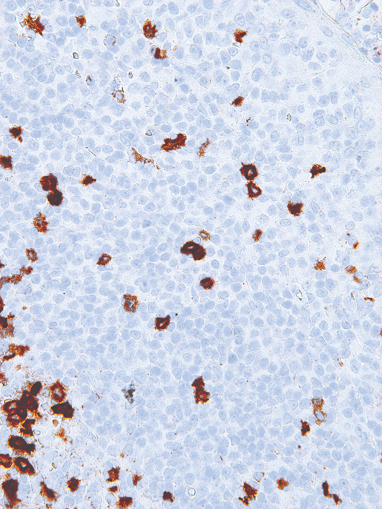
Figure 1 / 6

Click for expanded view
| Kit Specifications | |
| Minimum Urine Input | 11 mL |
| Maximum Urine Input | 30 mL |
| Size of RNA Purified | All sizes, including miRNA and small RNA (< 200 nt) |
| Elution Volume | 50 – 100 µL |
| Time to Complete 10 Purifications | 40 – 45 minutes |
| Average Yields* | Variable depending on the specimen |
*Please check page 5 of the product insert for the average yields and the common RNA quantification methods.
Storage Conditions and Product Stability
All buffers should be kept tightly sealed and stored at room temperature. This kit is stable for 2 years after the date of shipping. It is recommended to warm Lysis Buffer A for 20 minutes at 60°C if any salt precipitation is observed.
Important Note
Urine samples stored at -70°C, -20°C or at 4°C will develop some precipitation due to the aggregation of some of the highly abundant proteins in urine. Eliminating these precipitates using centrifugation or filtration may cause the loss of exosomes. Furthermore, these precipitates may affect the quality of the purified nucleic acid. We recommend the use of Norgen’s Urine Preservative when collecting urine samples, which is designed for the preservation of nucleic acids and proteins in fresh urine samples at ambient temperatures. The components of the Urine Preservative allow samples to be stored for over 2 years at room temperature with no detected degradation of urine DNA, RNA or proteins. Norgen’s Urine Preservative is available as a liquid format in Norgen’s Urine Preservative Single Dose Ampules, as well as in a dried format in Norgen’s Urine Collection and Preservation Tubes.
| Component | Cat. 59200 (50 preps) | Cat. 59300 (25 preps) | Cat. 59400 (15 preps) |
|---|---|---|---|
| Slurry E | 12.5 mL | 12.5 mL | 12.5 mL |
| ExoC Buffer | 8 mL | 30 mL | 50 mL |
| ExoR Buffer | 12 mL | 12 mL | 12 mL |
| Lysis Buffer A | 2 x 20 mL | 2 x 20 mL | 2 x 20 mL |
| Lysis Additive B | 2 mL | 2 mL | 2 mL |
| Wash Solution A | 2 x 18 mL | 18 mL | 18 mL |
| Elution Solution A | 2 x 6 mL | 6 mL | 6 mL |
| Mini Filter Spin Columns | 50 | 25 | 15 |
| Mini Spin Columns | 100 | 50 | 30 |
| Collection Tubes | 100 | 50 | 30 |
| Elution Tubes (1.7 mL) | 100 | 50 | 30 |
| Product Insert | 1 | 1 | 1 |

Permagen’s 32-tube PCR Separation rack features all of the same aspects of our MSR812 rack above for easy protocol transition for labs wishing to scale up to four PCR strips at a time, or up to 32 individual PCR tubes.
Features include solid aluminum alloy design with hard coat finish for years of trouble-free use, rubber feet to help prevent slipping on work bench, less tippy than common plastic products, and fast separations using any magnetic beads
MSR402
Maximum – .2 mL
Minimum – 30 µL
Permagen’s 32-tube PCR Separation rack features all of the same aspects of our MSR812 rack above for easy protocol transition for labs wishing to scale up to four PCR strips at a time, or up to 32 individual PCR tubes.

NH-bis(PEG2-propargyl) is a multi-branched linker with two terminal propargyl groups and an amino group. The propargyl groups reacts with azide-bearing compounds or biomolecules via copper catalyzed Click Chemistry to yield a stable triazole linkage. The amino group is reactive with carboxylic acids, activated NHS esters, and carbonyls. Reagent grade, for research purpose. Please contact us for GMP-grade inquiries.

NH-bis(PEG2-propargyl) is a multi-branched linker with two terminal propargyl groups and an amino group. The propargyl groups reacts with azide-bearing compounds or biomolecules via copper catalyzed Click Chemistry to yield a stable triazole linkage. The amino group is reactive with carboxylic acids, activated NHS esters, and carbonyls. Reagent grade, for research purpose. Please contact us for GMP-grade inquiries.
